Profliand
On this page, you find all documents, package deals, and flashcards offered by seller profliand.
- 69
- 0
- 5
Community
- Followers
- Following
1 Reviews received
74 items
DEVELOPMENT OF THE RESPIRATORY SYSTEM
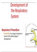
Development of the Respiratory System Respiratory Primordium • Primordium is an organ, structure or tissue in the earliest stage of development. • At the end of the 4th week IUL, respiratory primordium is indicated by a ventral outgrowth-the laryngo-tracheal groove (narrow cut) • It invaginates to form a pouch-like laryngotracheal diverticulum (blind tube), which is located ventral to the caudal part of the foregut NB/ Epithelium of the interna...
- Package deal
- Summary
- • 32 pages •
Development of the Respiratory System Respiratory Primordium • Primordium is an organ, structure or tissue in the earliest stage of development. • At the end of the 4th week IUL, respiratory primordium is indicated by a ventral outgrowth-the laryngo-tracheal groove (narrow cut) • It invaginates to form a pouch-like laryngotracheal diverticulum (blind tube), which is located ventral to the caudal part of the foregut NB/ Epithelium of the interna...
CARDIOVASCULAR SYSTEM DEVELOPMENT
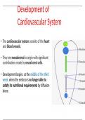
Development of Cardiovascular System • The cardiovascular system consists of the heart and blood vessels. • They are mesodermal in origin with significant contributions made by neural crest cells. • Development begins at the middle of the third week, when the embryo is no longer able to satisfy its nutritional requirements by diffusion alone. • Progenitor heart cells lie in the epiblast, immediately adjacent to the cranial end of the primitive streak. •...
- Summary
- • 45 pages •
Development of Cardiovascular System • The cardiovascular system consists of the heart and blood vessels. • They are mesodermal in origin with significant contributions made by neural crest cells. • Development begins at the middle of the third week, when the embryo is no longer able to satisfy its nutritional requirements by diffusion alone. • Progenitor heart cells lie in the epiblast, immediately adjacent to the cranial end of the primitive streak. •...
CARDIOVASCULAR SYSTEM ANATOMY
CARDIOVASCULAR SYSTEM Introduction to CVS • CVS is a closed system is the transport system in the body • It delivers oxygen, nutrients, hormones, and other important substances to cells in the body Components 1. The heart 2. Blood vessels 3. The Blood The Heart • Heart is a hollow muscular pumping organ • Shape- Pyramidal or conical. • Measurements- Length 12 cm, Width9 cm in adults • Weight-300 g in males; 250...
- Summary
- • 55 pages •
CARDIOVASCULAR SYSTEM Introduction to CVS • CVS is a closed system is the transport system in the body • It delivers oxygen, nutrients, hormones, and other important substances to cells in the body Components 1. The heart 2. Blood vessels 3. The Blood The Heart • Heart is a hollow muscular pumping organ • Shape- Pyramidal or conical. • Measurements- Length 12 cm, Width9 cm in adults • Weight-300 g in males; 250...
ANATOMY OF THE URETER
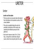
URETER Ureter Location and Description • The two ureters are muscular tubes that extend from the kidneys to the posterior surface of the urinary bladder . • The urine is propelled along the ureter by peristaltic contractions of the muscle coat, assisted by the filtration pressure of the glomeruli. • Each ureter measures about 10 in. (25 cm) long , having three constrictions along its course: where the renal pelvis joins the ureter, where it is kinked as it crosses the pe...
- Summary
- • 11 pages •
URETER Ureter Location and Description • The two ureters are muscular tubes that extend from the kidneys to the posterior surface of the urinary bladder . • The urine is propelled along the ureter by peristaltic contractions of the muscle coat, assisted by the filtration pressure of the glomeruli. • Each ureter measures about 10 in. (25 cm) long , having three constrictions along its course: where the renal pelvis joins the ureter, where it is kinked as it crosses the pe...
ANATOMY OF THE THYMUS GLAND
THYMUS THYMUS GLAND • Located behind/posterior to the manibrium sterni • Thymus gland has two parts:- • The cortex: that has densely packed T-lymphocytes • The thymus is equipped with a blood-thymus barrier, which is restricted to the cortex • Medulla has: Less densely parked T-lymphocytes It has hassal corpuscles It is enclosed by a higly vascularized tissue capsule Thymus under low magnification Cortex and medulla The cortex • It ...
- Summary
- • 8 pages •
THYMUS THYMUS GLAND • Located behind/posterior to the manibrium sterni • Thymus gland has two parts:- • The cortex: that has densely packed T-lymphocytes • The thymus is equipped with a blood-thymus barrier, which is restricted to the cortex • Medulla has: Less densely parked T-lymphocytes It has hassal corpuscles It is enclosed by a higly vascularized tissue capsule Thymus under low magnification Cortex and medulla The cortex • It ...
ANATOMY OF THE THORAX
Anatomy of the Thorax Thoracic wall Layers of the thoracic wall From superficial to deep; 1. Skin. 2. Superficial fascia. 3. Deep fascia. 5. Bones 4. Muscles. 5.Blood supply /innervation 5. Endothoracic fascia 6. Parietal pleura 1. The Skin -Is a thin skin -Hair distribution is variable and depends on the age, sex and race -Lines of cleavage lies horizontal 2. Superficial fascia -Is more dense on the posterior aspect of the chest to sustain the pressure of the body w...
- Package deal
- Summary
- • 42 pages •
Anatomy of the Thorax Thoracic wall Layers of the thoracic wall From superficial to deep; 1. Skin. 2. Superficial fascia. 3. Deep fascia. 5. Bones 4. Muscles. 5.Blood supply /innervation 5. Endothoracic fascia 6. Parietal pleura 1. The Skin -Is a thin skin -Hair distribution is variable and depends on the age, sex and race -Lines of cleavage lies horizontal 2. Superficial fascia -Is more dense on the posterior aspect of the chest to sustain the pressure of the body w...
anatomy of the testis
TESTIS INTRODUCTION • The testis is a male gonad. • It is homologous with the ovary in the female. • It is a mobile organ and lies in each half of the scrotal sac. • The functions of the testis include production of spermatozoa and secretion of testosterone (or dihydrotestosterone), a male hormone, responsible for the development and maintenance of the secondary sex characteristics of the mal SIZE • It measures approximately 4 X3 X 2.5 cm (length 4 cm,...
- Summary
- • 35 pages •
TESTIS INTRODUCTION • The testis is a male gonad. • It is homologous with the ovary in the female. • It is a mobile organ and lies in each half of the scrotal sac. • The functions of the testis include production of spermatozoa and secretion of testosterone (or dihydrotestosterone), a male hormone, responsible for the development and maintenance of the secondary sex characteristics of the mal SIZE • It measures approximately 4 X3 X 2.5 cm (length 4 cm,...
ANATOMY OF THE PERINIUM
PERINEUM • The perineum is traversed by • urethra and anal canal in the male, and • urethra, vagina, and anal canal in the female. • The surface features of the perineum • in male are penis, scrotum, and anal orifice, • in the female are vulva (female external genitalia) and anal orifice . • The median region between vaginal and anal orifices in female, containing perineal body is considered as gynecological perineum. Boundaries • The perineu...
- Summary
- • 103 pages •
PERINEUM • The perineum is traversed by • urethra and anal canal in the male, and • urethra, vagina, and anal canal in the female. • The surface features of the perineum • in male are penis, scrotum, and anal orifice, • in the female are vulva (female external genitalia) and anal orifice . • The median region between vaginal and anal orifices in female, containing perineal body is considered as gynecological perineum. Boundaries • The perineu...
ANATOMY OF THE PELVIS AND PERINIUM
PELVIC AND PERINEUM Introduction • The pelvis is the part of the trunk infero-posterior to the abdomen and is the area of transition between the trunk and the lower limbs. • The pelvic cavity is the inferior most part of the abdomino pelvic cavity. • Anatomically, the pelvis is the part of the body surrounded by the pelvic girdle (bony pelvis), part of the appendicular skeleton of the lower limb. Cont…. • The pelvis is subdivided into greater and lesser pelves....
- Summary
- • 54 pages •
PELVIC AND PERINEUM Introduction • The pelvis is the part of the trunk infero-posterior to the abdomen and is the area of transition between the trunk and the lower limbs. • The pelvic cavity is the inferior most part of the abdomino pelvic cavity. • Anatomically, the pelvis is the part of the body surrounded by the pelvic girdle (bony pelvis), part of the appendicular skeleton of the lower limb. Cont…. • The pelvis is subdivided into greater and lesser pelves....
ANATOMY OF THE PANCREASE
ANATOMY OF THE PANCREAS INTRODUCTION • The pancreas is a soft, finely lobulated, elongated exoendocrine gland. • The exocrine part secretes the pancreatic juice and the endocrine part secretes the hormones like insulin, etc. • The pancreatic juice helps in the digestion of lipids, carbohydrates, and proteins, whereas the pancreatic hormones maintain glucose homeostasis. Cont…. • The pancreas lies more or less horizontally on the posterior abdominal wall in the epiga...
- Summary
- • 32 pages •
ANATOMY OF THE PANCREAS INTRODUCTION • The pancreas is a soft, finely lobulated, elongated exoendocrine gland. • The exocrine part secretes the pancreatic juice and the endocrine part secretes the hormones like insulin, etc. • The pancreatic juice helps in the digestion of lipids, carbohydrates, and proteins, whereas the pancreatic hormones maintain glucose homeostasis. Cont…. • The pancreas lies more or less horizontally on the posterior abdominal wall in the epiga...

Driver's Ed: Chapter 7 Test Study Questions and Answers