Medical physiology breathing part 1: ventilation and work of breathing Van Duinen
Aims and components; airways and spirometry
The aim of breathing is to supply the cells in the body with oxygen and to get rid of the carbon
dioxide that is produced in the cellular metabolism. When we talk about breathing, we focus on
many components of breathing. While most of the time, we only think about the first component,
which is ventilation. Ventilation is the movement of air in and out of the lungs. It is the transport of
gasses in and out of the lungs. However, the other components are also part of breathing. These
parts are:
- Alveolar diffusion → diffusion of gases over the membranes. It is the
diffusion of gases (oxygen and carbon dioxide) between alveoli and blood.
- Perfusion of the lungs → without the pulmonary circulation, there is no
perfusion going on.
- How ventilation and perfusion are adjusted to each other. It is the tuning
of the ventilation and lung perfusion.
- Gas transport → the transport of oxygen to the tissues and carbon
dioxide back to the lungs. It is the transport of gasses in the blood.
- In the tissues there is also the diffusion of the gasses, the gas exchange
(or diffusion). It is the exchange of gas between blood and cells.
- In cells, we have cellular respiration, or metabolism. This is also part of
breathing. It is the use of oxygen and production of carbon dioxide in the
cells.
In this lecture, we look at the ventilation and the work of breathing (the mechanics
of ventilation).
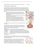
The anatomy of the airways. There is an upper respiratory system, which involves
the nasal cavity, the mouth, the pharynx and the larynx. The lower respiratory
system includes the trachea, bronchi and the right lung consisting of three lobes and
the left lung consisting of two lobes and the diaphragm.
We can split the bronchi up in the conducting zone and a
respiratory zone. The conducting zone is the zone in which the air is
conducted, but we can’t do much with the air when it is in this zone. So, it is
also called the anatomical dead space. When the air reaches the respiratory
zone, which consists of the smaller bronchioles and the alveoli, there the gas
exchange can take place. So, only the air that reaches this zone takes part in
the gas exchange.
There are capillary networks surrounding the alveoli and they are in the walls between the alveoli,
this makes that there is a large area available for the gas exchange. The gas exchange is between the
alveoli and the long capillaries.
When we do spirometry, we can look at the volumes that someone breathes.
The spirometer is filled with air, and it is connected to the participant. The top
of the spirometer is in a bucket of water. This means that if you suck in air, you
pull the chamber downwards and the pen will go upwards, to draw a
spirometer trace. In the spirogram, someone is breathing quietly in and out at
first. There is a maximal breath in and out, then normal breathing again, then a
,maximal breath out and in again, then we see a maximal breath in followed by a maximal breath out
and normal breathing again. These different volumes have names, we can add volumes together and
then you get capacities. When someone is breathing quietly, we talk about the tidal volume (VT). This
tidal volume in a healthy person is, on average, about 500 ml per breath. If you breath in extra air,
the amount of air that we can breathe in extra is the inspiratory reserve volume (IRV), this is about 3
litres. If we add the tidal volume to the inspiratory reserve volume, we have the inspiratory capacity.
If someone breathes out extra after normal breathing. This extra air that we can breathe out is called
the expiratory reserve volume (ERV). If someone breathes out completely, there is still some air in
the lungs left, this is called the residual volume (RV). This volume is always in the lungs. Normally, if
we start breathing in, we also have the expiratory reserve volume in our lungs. When we add the
residual volume and the expiratory reserve volume, we get the functional residual capacity. This
means that when you start breathing normally, you already have 2/2,5 litres of air in your lungs. So,
we start breathing at the functional residual capacity. The amount that you can breathe in maximally
and breath out maximally, if we add this, you get the vital capacity. This is the maximal amount of air
that you can breathe in and breath out. If we add the residual volume to the vital capacity, you have
the total lung capacity.
Functional residual capacity (amount of air you have in the lungs when you start breathing in) → FRC = ERV + RV
Inspiratory capacity → IC = VT + IRV
Vital capacity → VC = IC + ERV
Total lung capacity → TLC = VC + RV
Note → sometimes you do a test in which someone needs to breath out maximally at once, then we
call it the forced vital capacity. Then it will be the FVC.
Ventilation
Ventilation is the movement of air in and out of the lungs. How can
we make sure that the air moves into the lungs and out again? We
do this by using our muscles of inspiration. We contract the
diaphragm so that it flattens. When this happens, we enlarge the thoracic volume. If we enlarge the
thorax and if it were a closed space, then the pressure multiplied with the volume stays the same.
This is a constant → Boyle’s law (PxV = constant). If we enlarge the volume, this means that the
pressure goes down. Air flows from a high pressure to a low pressure. So, if we lower the pressure
inside the lungs, the air will flow from the outside into the lungs. If we then relax the muscles of
inspiration, the thorax and lungs will go back to their resting position and the volume decreases and
the air will be pushed out of the lungs. When we ventilate, we create pressure changes in the lung.
These pressure differences cause the flow of air. We use the diaphragm for inspiration, but we also
use intercostal muscles (muscles between the ribs). We have two layers of these muscles. The
external intercostal muscles are the muscles that lie on the outside. When we contract these
muscles, we lift the ribs up and out. We use those muscles for inspiration. So, for inspiration we
contract the external intercostal muscles and the diaphragm, enlarging the thoracic cavity and then
air flows in. For expiration, we relax those muscles, the lungs go back to the initial position and the
air is pushed out. In black is our lung, the green is our thoracic cavity. There is a space between the
lungs and the cavity, this space is called the pleural space. When we contract the muscles of
inspiration, we enlarge the thorax. The volume within the pleural space increases
in size as well, this means that the pressure in the pleural space becomes lower,
and it pulls on the lungs and the lungs will become expanded. The amount that
the lung expands depends on how compliant the lung is. If the lung is very stiff,
then we need to create a large pressure difference to be able to expand the lung.
If the lung is very floppy, only a small pressure difference is needed to expand the
lung. Due to the expansion, the pressure inside the alveoli goes down, and then
,air starts to flow. The amount of air that flows depends on the resistance. The resistance depends on
how narrow the bronchiole is and how much air is flowing. The more the molecules in the air bounce
against each other, the more resistance there is. This happens more if the bronchiole is very narrow,
constricted. When there is bronco dilation, they are wider and then the resistance is lower, and the
air can flow easier.
Forces that need to be overcome when we are breathing (ventilating) → We have to overcome the
elastic forces (‘static forces'), or better said, the retraction forces and we have to overcome the
resistance forces (‘dynamic’), there is both airway resistance and tissue resistance (airway has a way
bigger effect than tissue resistance in healthy people).
The retraction forces of the lung and thorax wall. In the picture is the ribcage, lung, parietal pleura
(lining the thorax), pulmonary pleura (lining the lungs). In between the pleura is the pleural space.
The heart is in its pericardial sac. The favourite position of the thorax would be nice and wide. It can’t
be because if we look at the two membranes, there is a little layer of fluid in between which pulls
those membranes together. Even though the thorax wants to be big, the lungs want to be small, but
they can’t because of the layer of fluid between the pleura. So, the thorax is pulling to the outside,
the lungs are pulling to the inside. This makes that the pressure in the pleural space is lower than the
pressure in the outside world and lower than the pressure inside the lungs. The outside air pressure
is about 760 mm mercury. Inside the lungs, if there is no movement of air, the pressure is the same.
These values are very difficult to work with. So, usually we say for these cases when we look at
changes in the breathing, we pretend that the outside air pressure is 0. The pressure in the lungs is
also 0. Then the pressure in the space between the pleura is lower than the pressure in the lungs and
the outside pressure. If we pretend that the pressure in the lungs and the outside air are 0, this
means that the pressure in the pleural space becomes negative.
Now, we look at pressures over the wall. The pressure over the lung
wall (Pl) is the pressure inside the alveoli (Palv) – the pressure in the
pleural space (Ppl). If we are not breathing, the pressure in the alveoli is
0, the pressure in the pleural space was negative, which means that the
pressure over the wall is positive. It is important that this pressure is
positive, because as long as it is positive it pushes the alveoli open. If it
were to become negative, this means that the pressure in the pleural
space is bigger than the pressure in the alveoli, then the alveoli and bronchiole would be pushed
closed. We can also look at the pressure over the thoracic wall (chest wall), this is the pressure inside
the pleural space (Ppl) – the pressure in the outside air (Pb). As the pressure in the pleural space was
negative, and the pressure outside is 0, the pressure over the thoracic wall is negative. If we look at
the pressure over the two membranes (Pl + th) = pressure inside the alveoli (Palv) – the pressure of the
outside air (Pb). If we are not breathing, these pressures are the same, so the pressure over the lung
and thorax together will be 0. So, if we look at these transmural pressures, you always take the
pressure on the inside (Pin) – the pressure on the outside (Pout). How can you measure these
different pressures? How can you measure what the pressure inside the alveoli/pleural space etc. is?
Static (or relaxation) volume-pressure relationship (curve). The pressures
on the x-axis are transmural pressures. We have three transmural
pressures, namely, the pressure over the lung wall (Palv – Ppl) is positive at
FRC. The pressure over the thorax wall at FRC (the start of inspiration), this
is negative because there is a negative pressure in the pleural space, if you
subtract the pressure of the outside air, it is a negative pressure. The
pressure over the lung and the thorax together is the alveolar pressure –
the outside air pressure. At FRC (this is an individual who has 3 litres of air
inside the lung at the start of inspiration), what you normally do is breath
, in some air. As this is a static volume-pressure curve, they let some air into the lungs and then
measure the pressures (repeat this a couple of times and then draw a line). This means that at a
point a bit above the red circle, there is extra air in the alveoli compared to the resting situation and
therefore we have a higher pressure inside the alveoli compared to the outside air. That is why it
goes to the positive side. Most important is that at FRC, the lungs and thorax are in an equilibrium.
Meaning that the thorax wants to be big, so it is pulling, the lungs want to be small, so they are
pulling, but at the moment of the red circle they are pulling equally strong in different directions. The
steepness of this curve is determined by the compliance of the lung and thorax.
The compliance is the volume change that occurs with a certain pressure change. It is the difference
in volume divided by the difference in pressure (ΔV/ΔP). We can determine this from the static (or
relaxation) volume-pressure curve. The steepness of the static V/P-curve gives us information about
the compliance of the lungs. If it is easy to expend the lungs, it will go steep. If it is hard to expand
the lungs, the slope will be less steep.
The slope in the static volume-pressure curve is determined by the compliance of the lungs. What
makes that those lungs want to be small? First of all, this is determined by the number of elastin- and
collagen fibres and by the surface tension of the alveoli. First, we will look at the effect of surface
tension, as for the elastin- and collagen fibres, we start off with a certain
amount of elastin and collagen. Throughout life you lose elastin fibres and
therefore the tissue becomes stiffer. However, other changes may cause that
the lungs become sloppier. We focus on the surface tension of the alveoli. We
see that if we have the alveoli, there is a little layer of fluid along the
membranes. This layer wants to pull together to become a drip of fluid. This
means that there is a certain amount of force pulling those alveoli together. If
we see this in each alveolus, this makes that the whole lung wants to be small.
This means that if we would fill the lungs up with water and we look at the
normal pressure volume curve (white dots) in which the alveoli are filled
with air, we see that if we fill it up with water, the surface tension goes
away because it is all water. Then it is very easy to expend the lungs, we
only need small pressure changes to get a large volume change. The result
of surface tension is that we have a strong retraction force of lung in total,
and we could get collapse of smaller alveoli into bigger ones. This is because
we have the law of Laplace, which tells us that the pressure with which
those alveoli want to get together, is two times the surface tension divided
by r (radius of the alveolus. Law of Laplace: P = 2T/r. if the surface tension is
in principle the same between large and small alveoli, this means that the
pressure of going together is bigger in alveoli with a smaller radius. In the
figure you see that the smaller alveolus would empty itself into the bigger one as the pressure to
become smaller is way bigger compared to the big alveolus. Luckily, this doesn’t happen because we
have surfactant producing cells. Surfactant decreases the surface tension. We have a similar number
of surfactants producing cells in all alveoli. Because of this, in the smaller alveoli there are relatively
more surfactant producing cells, and thus there is relatively more surfactant compared to the larger
alveoli. So, we have a bigger reduction in surface tension in smaller alveoli, and thus the smaller
alveoli won’t empty in the bigger alveoli.
In the static volume-pressure curve, we see the resting respiratory level (FRC). We can breathe in (to
the upper part of the line) and out (to the lower part of the line). At FRC level, the thorax wants to be
big (there is a force from the thorax pulling to the outside), and the lungs want to be small (there is a
force from the lungs pulling to the inside). At FRC, these forces are equally big but in the opposite