N164: Managing Care of Adults III
Intracranial Pressure Regulation Problems and Acute Head Injury Ch. 61 W3
Intracranial Regulation • Components of the Brain
o Three essential volume components:
1. Brain tissue (1,400g approx.)
2. Blood (75mL approx.)
3. CSF (75mL approx.)
• Brain Injury Categorized in 2 Phases
1. Primary Injury
• Occurs at the initial time of an injury (e.g., impact of car accident, blunt-force trauma).
• Results in displacement, bruising, or damage to any cranial component (brain tissue, blood,
CSF).
2. Secondary Injury
• The resulting hypoxia, ischemia, hypotension, edema, or increased ICP that follows the
primary injury.
• Can occur several hours to days after the initial injury.
• It is the modifiable concern when managing brain injury.
o Management of the patient with an acute intracranial problem must include managing secondary
injury and preventing increased ICP.
Normal Intracranial Pressure " Intracranial pressure (ICP) = the hydrostatic force measured in the brain CSF compartment
Normal ICP = 5–15 mmHg o Under normal conditions in which intracranial volume stays relatively constant, the balance
Elevated ICP > 20 mmHg among the 3 components (brain tissue, blood, CSF) maintains ICP.
for more than 15 minutes o We can measure ICP in the ventricles, subarachnoid space, subdural space, epidural space, or
brain tissue with a pressure transducer.
• Intracranial Pressure Regulation: Factors that influence ICP
o Arterial pressure (Hypertension)
o Venous pressure (Return flow)
o Intra-abdominal Pressure (Ascites)
o Intra-thoracic pressure (Coughing)
o Posture (Neck flexed)
o Temperature (Hyperthermia)
o Blood gases (especially CO2 levels)
* The degree to which these factors increase or decrease the ICP depends on the brain’s ability to
adapt to changes.
• Monro-Kellie Doctrine/Hypothesis
" Only applicable if skull is CLOSED
o States that the 3 components must stay at a relatively constant volume within the closed skull
o If the volume of any 1 of the 3 components increases within the skull and the volume from
another component is displaced, the total intracranial volume will not change. This hypothesis is
applicable only in situations in which the skull is closed. The hypothesis does not apply in
persons with displaced skull fractures or craniectomy (removal of part of the skull).
o Dictates: If one component increases, another must decrease to maintain normal compensatory
adaptations (ICP)
• Changes in CSF volume
• Changes in intracranial blood volume
• Changes in tissue brain volume
o When the ability to compensate is limited, we see change in patient condition
• If volume increase continues, ICP rises, leading to decompensation, which leads to
compression and potential herniation of brain tissues.
,Cerebral Blood Flow " CBF = The amount of blood in milliliters passing through 100 g of brain tissue in 1 minute
o The global CBF is about 50 mL/min/100 g of brain tissue.
o Maintaining blood flow to the brain is critical because the brain requires a constant supply of O2
and glucose. The brain uses 20% of the body’s O2 and 25% of its glucose.
• Autoregulation of CBF
o Automatic adjustment in diameter of cerebral blood vessels
o Ensures consistent CBF
o Only effective if mean arterial pressure (MAP) 70 to 150 mm Hg
o CPP = MAP – ICP
o Need to keep CPP > 65 mm Hg
• Hypoxia if < 60 mm Hg
• Necrosis if < 50 mm Hg
• Stages of Increased ICP
o Stage 1: Total compensation
o Stage 2: ↓ Compensation; Risk for ↑ICP
o Stage 3: Failing compensation; Clinical manifestations of increased ICP (Cushing’s triad)
o Stage 4: Herniation imminent leading to death
Increased Intracranial Pressure • Causes
o Acute Head Injuries: May be open or closed
• Closed Head Injuries:
◦ Subdural/Epidural/Subarachnoid Bleeds
◦ Interventricular Bleed
◦ Brain Tumors
◦ Cerebral Edema: Vasogenic; Cytotoxic; Interstitial
• Open Head Injuries:
◦ Gunshot Wounds/Weapons
◦ War Traumas
• Idiopathic (unknown Source)
• Infections (Meningitis)
• Aneurysms
• Motor Vehicle Accidents
• Sports related Injuries
• Assaults
• Facts about TBI
o TBI’s more common in younger males
o All TBI’s have a risk of poor outcome if not managed well.
o Death can occur at three points following TBI
• Immediately after the injury
• Within a few hours after injury
• About a few weeks after injury
, • Clinical Manifestations
o Ocular Signs → Compression of cranial nerve (CN) III oculomotor nerve
• Ipsilateral pupil dilation (same side as the mass lesion)
• Sluggish or no response to light
• Inability to move eye upward, adduct
• Eyelid ptosis
• Fixed, unilateral, dilated pupil is considered a neurologic emergency
o Other Cranial Nerves
• Optic (CN II), trochlear (CN IV), and abducens (CN VI) nerves
• Diplopia, blurred vision, EOM changes
o Decrease in Motor Function
• Contralateral hemiparesis/hemiplegia
• Decerebrate posturing (extensor) → Indicates more serious damage
◦ The arms are stiffly extended, adducted, and hyperpronated. There is hyperextension
of the legs with plantar flexion of the feet.
• Decorticate posturing (flexor)
◦ Consists of internal rotation and adduction of the arms with flexion of the elbows,
wrists, and fingers. It is a result of interruption of voluntary motor tracts in the
cerebral cortex. Extension of the legs may be seen.
o Change in Level of Consciousness
• Flattening of affect associated with coma
o Changes in Vital Signs
• Cushing’s Triad
1. Systolic HTN w/widening pulse pressure
2. Bradycardia with a full and bounding pulse
3. Irregular respirations
• Change in body temperature
o Headache
• Often continuous
• Nocturnal/Morning headache
o Vomiting
• Not preceded by nausea (aka unexpected vomiting)
• Projectile vomiting may occur
• Complications
o Inadequate cerebral perfusion
o Cerebral herniation
• Tentorial herniation (central herniation)
◦ Occurs when a mass lesion in the
cerebrum forces the brain to
herniate downward through the
opening created by the brainstem
(the foramen magnum).
• Uncal herniation
◦ Lateral and downward herniation
• Cingulate herniation
◦ Lateral displacement of brain tissue
beneath the falx cerebri
o To help understand cerebral herniation, we are going to describe 2 important structures in the
brain. The falx cerebri is a thin wall of dura that folds down between the cortex, separating the 2
cerebral hemispheres. The tentorium cerebelli is a rigid fold of dura that separates the cerebral
hemispheres from the cerebellum. It is called the tentorium (meaning tent) because it forms a
tent-like cover over the cerebellum.
• Diagnostic Studies
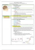
o Cerebral Angiography (Uses Contrast Media–CT w/Contrast)
• Check Renal Function
• Check Allergies to Iodine & Shellfish
← Normal brain blood flow
← Presence of cerebral aneurysm that can cause decease cerebral
blood flow may lead to increase ICP