N164: Managing Care of Adults III
Respiratory Failure Ch. 28, 30, 32 W2
Gas Exchange Unit
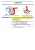
Respiratory System Overview " The primary purpose of the respiratory system is gas exchange. This involves the transfer of oxygen
(O2) and carbon dioxide (CO2) between the atmosphere and blood. While adequate perfusion is
needed to distribute O2 to the body tissues, adequate oxygenation depends on a healthy,
functioning respiratory system.
o Heart → Drives hemoglobin to cells
o Lungs → Oxygenate and ventilate that hemoglobin
To oxygenate and ventilate the hemoglobin, three things must work well:
1. Brainstem (medulla) → Controls rate and character of respirations
2. Muscles/Diaphragm → Controls ventilation
3. Alveolar capillary membrane → Controls oxygenation (respiration)
O2 and CO2 move back and forth across the alveolar-capillary membrane by diffusion
o Direction of movement → from the area of higher concentration to the area of lower
concentration
o Thus, O2 moves from alveolar gas (atmospheric air) into the arterial blood and CO 2 from the
arterial blood into the alveolar gas.
o Diffusion continues until equilibrium is reached.
, O2-Hemaglobin Disassociation
Curve
Rapid Sequence Intubation (RSI) 1. Preparation: IV, Suction, NRB, BVM, Laryngoscope, ET tubes, CO2 detector, ET tube stylets, Bougie,
Bite Block/Tape, Crash cart, RT
2. Pre-Oxygenation: 5 mins 100% O2 via NRB, End-Tidal CO2 (35-45%) Succinylcholine
3. Pretreatment & Induction: Etomidate, Ketamine, Propofol, Fentanyl Malignant Hyperthermia
o Hypotension → NO Etomidate or Propofol • Tachycardia
o Epilepsy → NO Etomidate • Elevated Temp
4. Paralytic Medications: Succinylcholine, Vecuronium, Rocuronium • Elevated EtCO2
5. Placement: ET size, GlideScope Tx: Dantrolene
6. Placement Verification: Calorimetric CO2 detector, bilateral breath sounds, chest wall rise, No gastric
sounds, chest x-ray, Capnography, O2 readings
7. Postintubation Management: Sedation/analgesia, DOPE, suctioning
Rapid sequence induction (RSI) is the rapid, concurrent administration of both a sedative and a
paralytic drug during emergency airway management to induce unconsciousness for intubation. It
decreases the risks for aspiration and injury. RSI is not indicated in patients who are in cardiac arrest
or have a known difficult airway. The patient receives a sedative-hypnotic-amnesic (e.g., propofol,
etomidate) to induce unconsciousness and a rapid-onset opioid (e.g., fentanyl) to blunt the pain of the
procedure. This is followed with a drug (e.g., rocuronium) to produce skeletal muscle paralysis.
During intubation, monitor vital signs, including heart rate and rhythm, signs of visible chest
movement, BP, and mean arterial pressure (MAP). Monitor O2 status with pulse oximetry.
Immediately inform the team if the SpO2 is less than 92%. Monitor the patient’s tolerance to the
procedure. Follow agency policy about RSI drug administration.
Immediately after intubation, inflate the cuff on the ET tube. Help confirm placement of the ET tube
while continuing to manually ventilate the patient using the BVM with 100% O2. Auscultate the lungs
for bilateral breath sounds and the epigastrium for the absence of air sounds. Observe for symmetric
chest wall movement. SpO2 should be stable or improve. Use an EtCO2 detector to help confirm
placement by noting the presence of exhaled CO2 from the lungs. A portable EtCO2 monitor can help
confirm correct placement of the tube. A steadily rising CO2 value and the presence of a waveform
confirms correct placement. Or place a colorimetric CO2 detector between the BVM and ET tube and
look for a color change (indicating the presence of CO2) or a number. At least 5 or 6 exhalations with
a consistent CO2 level must occur to confirm tube placement in the trachea.
If the findings support ET tube placement, help secure the ET per agency policy. Connect the ET tube
to a ventilator and closed suctioning system. Assess the need to suction the ET tube and pharynx.
Insert a bite block, if needed. Secure it separately from the ET tube to the patient’s face. This will help
prevent the patient from biting the ET and preventing O2 delivery.
Obtain a chest x-ray to confirm tube location (2 to 3 cm above the carina in the adult). This position
allows the patient to move the neck without moving the tube or causing it to enter the right mainstem
bronchus. Once positioning is confirmed with x-ray, record and mark the position of the oral ET tube