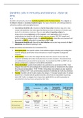
Summary Advanced Immunology Janeway test 2) Dendritic cells in immunity and tolerance
97 views 1 purchase
Course
Advanced Immunology (5234ADIM6Y)
Institution
Universiteit Van Amsterdam (UvA)
Book
Janeway\'s Immunobiology
This is a small summary for the course advanced immunology from the master biomedical sciences at the UvA. It includes all the information you need for one of the 9 Janeway tests during this course. Look out for the bundle, because that's a lot cheaper!
Janeways Immunobiology 9th Edition by Kenneth Murphy; Casey Weaver |
Janeways Immunobiology 9th Edition by Kenneth Murphy; Casey Weaver |
Janeways Immunobiology 9th Edition by Kenneth Murphy; Casey Weaver |
All for this textbook (19)
Written for
Universiteit van Amsterdam (UvA)
Biomedische wetenschappen
Advanced Immunology (5234ADIM6Y)
All documents for this subject (9)
Seller
Follow
nadinevankleef
Reviews received
Content preview
Dendritic cells in immunity and tolerance – Ester de
Jong
9.8
Dendritic cells primarily arise from myeloid progenitors within the bone marrow. They migrate via
the blood to tissues or secondary lymphoid organs. Two types of dendritic cells (distinguishable by
cell surface markers and transcription factors):
1. Conventional dendritic cells: Abundant at barrier tissue sites (intestines, lung, skin, close
contact with epithelia). Also solid organs like kidney and heart. In absence of infection: Low
levels of co-stimulatory molecules. They are very active in ingesting antigens by
phagocytosis using complement and FC receptors, and C type lectins which recognise
carbohydrates (on dendritic cells they include mannose receptor. DEC 205, langerin and
Dectin 1). Uptake of antigens directs it to endocytic pathway, where they are processed and
presented on MHC II molecules for recognition by CD4 T cells.
2. Plasmacytoid dendritic cell: Primarily for viral infection and secrete large amounts of class I
interferons, less efficient in priming T cells.
Antigen processing and presentation by (conventional) DCs:
Macropinocytosis: Non specific uptake of extracellular antigens including surrounding fluid.
Used for taking up bacteria that have mechanisms to evade recognition by phagocytic
receptors.
Viral infection: Occurs when the antigen directly enters the cytosol. Viruses enter the
cytoplasm by binding cell surface molecules that act as entry receptors. Viral proteins that
are synthesised are processed in the proteasome presented into MHC I so CD8 T cell can
activate and become a cytotoxic T cell to kill the infected DC.
Cross presentation: Uptake of extracellular virus particles to be presented on MHC I. Viral
antigens that enter the cell via phagocytic or endocytic vesicles are transferred to the cytosol
for proteasomal degradation ER MHC I. Extracellular viral antigens are also loaded on
MHC II so effector CD4 T cells can stimulate production of antibodies by B cells (by cytokines).
Transfer: Dendritic cells in the skin capture the antigens transport lymph nodes antigen
transferred to resident CD8-alfa positive dendritic cells (these are responsible for priming
naive CD8 T cells). So antigens from viruses that rapidly kill DCs can still be presented to
uninfected DCs that have been activated by their TLRs.
, 9-10
DCs capture pathogens by phagocytic receptors/macropinocytosis activate
responses through pattern recognition receptors such as TLRs. In humans,
conventional DCs express all known TLRs except for TLR-9.
Plasmacytoid DCs express TLR-9, TLR-1 and TLR-7.
Several of the phagocytic receptors also provide maturation signals:
DC-SIGN which binds mannose and fucose present on pathogens.
Dectin-1 which recognises beta-1,3-linked glucans on fungal cell walls.
Other receptors that bind pathogens (complement & phagocytic
receptors mannose receptor) also help DC activation.
TLR signalling results in alteration chemokine receptors on DCs. This change is
called licensing they will now activate T cells. The CCR7 receptor is now
activated, this makes DCs sensitive to CCL21 produced by lymphoid tissue. They
migrate directly into the T cell zones from the marginal sinus.
CCL21 signalling through CCR7 also results in expression of MHC I and
MHC II while they cannot engulf antigens anymore.
There is also expression of costimulatory molecules on their surface,
B7.1 (CD80) and B7.2 (CD86). They deliver costimulatory signals to naive
T cells.
They also express high levels of adhesion molecules DC-SIGN and
secrete chemokine CCL19 attracts naive T cells.
DCs also present self peptides, T cell receptors (normally) do not recognise
these. Besides, the DC with self peptides does not express co-stimulatory
molecules.
Not only peptides activate DC activation:
Bacterial/viral DNA containing unmethylated CpG motifs recognised by TLR-9
(intracellular). This activates NfkappaB and MAPK pathways pro inflammatory cytokines
(IL-6, IL-12, IL-18) and expression co stimulatory molecules on DCs.
HSP(heat shock protein) from bacteria antigen presentation.
Double stranded RNA from viruses antigen presentation.
For costimulatory molecule expression you need bacteria/bacterial components known as adjuvants.
9-15
Activation of naive T cells consists of 3 signals:
1. Interaction of a peptide & MHC complex with the T cell receptor.
2. Co-stimulatory signals that promote survival and expansion of T cells
3. Cytokines that direct T cell differentiation into one of the effector T cells.
Additional signals include Notch ligands contribute to effector differentiation.
Co-stimulatory molecules are B7 molecules (CD80 & CD86). The receptor for B7 on the T cell is CD28.
B7 deficiency or blockage inhibit T cell response.
The benefits of buying summaries with Stuvia:
Guaranteed quality through customer reviews
Stuvia customers have reviewed more than 700,000 summaries. This how you know that you are buying the best documents.
Quick and easy check-out
You can quickly pay through credit card or Stuvia-credit for the summaries. There is no membership needed.
Focus on what matters
Your fellow students write the study notes themselves, which is why the documents are always reliable and up-to-date. This ensures you quickly get to the core!
Frequently asked questions
What do I get when I buy this document?
You get a PDF, available immediately after your purchase. The purchased document is accessible anytime, anywhere and indefinitely through your profile.
Satisfaction guarantee: how does it work?
Our satisfaction guarantee ensures that you always find a study document that suits you well. You fill out a form, and our customer service team takes care of the rest.
Who am I buying these notes from?
Stuvia is a marketplace, so you are not buying this document from us, but from seller nadinevankleef. Stuvia facilitates payment to the seller.
Will I be stuck with a subscription?
No, you only buy these notes for $6.11. You're not tied to anything after your purchase.