Anatomy of the pericardium, heart chambres, cardiac skeleton, coronary vasculature, Koch's Triangle, cardiac contraction, conduction system of the heart, innervation, ascending aorta, pulmonary trunk. Overview of the foetal circulation, and microanatomy of arteries and veins.
Anatomy of the Heart
The mediastinum is the central partition of the thoracic cavity,
going from the sternum to the bodies of vertebrae, and from the
superior thoracic aperture to the diaphragm. It separates the two
pleural cavities, and contains the thymus, pericardial sac, heart,
trachea, major arteries and veins. Serves as passageway for the
oesophagus, thoracic duct (largest lymphatic vessel), and various
nerves.
• A transverse plane going from sternal angle to intervertebral
disc T4-T5 divides the mediastinum into superior mediastinum
and inferior mediastinum.
• The inferior mediastinum is further subdivided into: anterior
mediastinum, the region between the pericardial sac and the
diaphragm, posterior mediastinum, between pericardial sac
and vertebral bodies, and middle mediastinum, which includes
the pericardial sac. In fact it contains the pericardium, heart,
origins of great vessels, smaller vessels and various nerves.
THE PERICARDIUM
The pericardium is a fibrous sac surrounding heart and roots of
great vessels.
• Fibrous pericardium, a outer, cone-shaped, bag of connective
tissue. Its apex is continuous with the adventitia of grater vessels, while the base is attached to the
central tendon of the diaphragm. Anteriorly it is attached to the posterior surface of the sternum by
sterno-pericardial ligaments. These attachments maintain heart position and limit cardiac distension.
Phrenic nerves pass through the fibrous pericardium to reach the diaphragm, innervating both. Similarly
pericardiaco-phrenic vessels supply both.
• Serous pericardium, thin layer divided into parietal layer, which lines the fibrous, and visceral layer
(epicardium), which adheres the heart. They are continuous at the root of great vessels (reflection
zones), aorta, pulmonary trunk, superior and inferior vena cava and pulmonary veins. They are
otherwise divided by a narrow space, the pericardial cavity, filled with fluid, which allows heart
movement. The reflection zones onto the pulmonary veins forms the oblique pericardial sinus. The site
separating the reflection zones of arteries and those of veins is the transverse pericardial sinus.
Blood supply to pericardium occurs through branches of internal thoracic, pericardiaco-phrenic, musculo-
phrenic and inferior phrenic arteries, and thoracic aorta. Veins draining the pericardium enter the azygos
system, internal thoracic and superior phrenic veins.
Fibrous pericardium and the parietal layer of serous pericardium are supplied by phrenic nerves. The
visceral layer of serous pericardium instead is innervated by branches of sympathetic trunks and vagus
nerves.
1
, sabato 2 marzo 2019
THE HEART
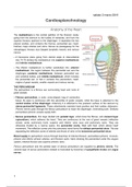
The heart is a pyramidal shaped
organ. The base of the heart (1) is
quadrilateral and directed posteriorly. It
consists of the left atrium, small portion
of right atrium, proximal parts of
superior and inferior venae cavae and
pulmunary veins. It is fixed posteriorly
to the pericardial wall, opposite to
vertebral bodies T5 to T8. The
oesophagus lies posterior to the base.
The apex of the heart (2) projects
forward, downward and to the left. It
includes the infero-lateral part of the
left ventricle and is positioned deep to
the 5th intercostal space, 8-9 cm from
the mid-sternal line.
The anterior surface of the heart (2)
mostly consists of the right ventricle,
with small portions of right atrium and
left ventricle.
The heart rests on the diaphragmatic
surface (3), which extends from the
base to the apex, divided from the
base by the coronary sinus. It consists
of the left ventricle and a small portion
of right ventricle, separated by the
posterior inter-ventricular
groove.
The left pulmonary surface
faces left lung and consists of
left ventricle and portion of left
atrium.
The right pulmonary surface
faces right lung and consists
of right atrium.
They are also referred to as
left and right margins.
The inferior margin is the
edge between anterior and
diaphragmatic surfaces.
The obtuse margin separates
anterior and left pulmonary
surfaces.
2
The benefits of buying summaries with Stuvia:
Guaranteed quality through customer reviews
Stuvia customers have reviewed more than 700,000 summaries. This how you know that you are buying the best documents.
Quick and easy check-out
You can quickly pay through credit card or Stuvia-credit for the summaries. There is no membership needed.
Focus on what matters
Your fellow students write the study notes themselves, which is why the documents are always reliable and up-to-date. This ensures you quickly get to the core!
Frequently asked questions
What do I get when I buy this document?
You get a PDF, available immediately after your purchase. The purchased document is accessible anytime, anywhere and indefinitely through your profile.
Satisfaction guarantee: how does it work?
Our satisfaction guarantee ensures that you always find a study document that suits you well. You fill out a form, and our customer service team takes care of the rest.
Who am I buying these notes from?
Stuvia is a marketplace, so you are not buying this document from us, but from seller Greta96. Stuvia facilitates payment to the seller.
Will I be stuck with a subscription?
No, you only buy these notes for $3.49. You're not tied to anything after your purchase.