Anatomy of the oral cavity, tongue, teeth, peritoneum, stomach, small intestine, large intestine, liver, gallbladder and spleen. Microanatomy of GI mucosa, liver, gallbladder and spleen.
The Digestive System
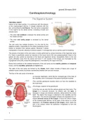
THE ORAL CAVITY
Inferior to the nasal cavities, it is continuous with the pharynx
at the oropharyngeal isthmus. It merges anteriorly with the
lips, surrounding the oral fissure. The oral cavity is separated
in two regions by the dental arches, consisting of teeth and
alveolar bone:
• The outer oral vestibule is between the dental arches and
the cheeks and lips.
• The inner oral cavity proper is enclosed by the dental
arches.
The oral cavity has multiple functions. It is the inlet for the
digestive system, responsible for the initial processing of food
thanks to secretion from salivary glands. Moreover it allows
manipulation of sounds, allowing to produce speech. In emergencies can as well be used for breathing.
The sensory innervation of the oral cavity is mainly performed by various branches of the trigeminal nerve
[V]. Taste is specifically carried by branches of the facial nerve [VII], and parasympathetic fibres to the
glands are also carried by branches of the facial and trigeminal nerves. Sympathetic fibres to the oral
cavity ultimately come from the spinal cord level T1. All muscles of the tongue are innervated by the
hypoglossal nerve [XII], except the palatoglossus, innervated by the vagus nerve [X].
Bones that contribute to the skeletal framework of the oral cavity are the maxilla, palatine and temporal
bones, and the mandible, sphenoid and hyoid bone.
The walls of the oral cavity are formed by the cheeks. Each cheek consists of fascia and a layer of
skeletal muscle (mainly buccinator) sandwiched between skin and oral mucosa.
The floor of the oral cavity is formed by:
•A muscular diaphragm, which fills the U-shaped gap of the body of
the mandible, and is composed of the paired mylohyoid muscles.
•Two cord-like genohyoid muscles which run from the mandible to
the hyoid bone.
•The tongue, superior to the geniohyoid.
In the floor we can also find the salivary glands and their ducts. The
tongue is a muscular structure. Its anterior part, the apex, is
triangular in shape and directed anteriorly, sitting behind the incisor
teeth. The root of the tongue is attached to the mandible and the
hyoid bone. The oral and pharyngeal surfaces are separated by a V-
shaped terminal sulcus of the tongue. It forms the inferior margin
of the oropharyngeal isthmus. At the apex of the sulcus is a small
depression, the foramen caecum of the tongue, which marks the
site in the embryo where the epithelium invaginated to form the
thryoid gland.
1
, giovedì 28 marzo 2019
The superior surface of the oral part of the tongue is covered by hundreds of papillae. These are
responsible for increasing the surface area. All except filiform papillae have taste buds on their surface.
• The filiform papillae are small cone-shaped projections of mucosa, ending in one or more points.
• The fungiform papillae are rounded in shape and larger, tend to be concentrated along the margins of
the tongue.
• The vallate papillae are the largest, cylindrical invaginations of the surface, immediately anterior to the
terminal sulcus.
• The foliate papillae are linear folds of the mucosa on the sides of the tongue, near the terminal sulcus.
The inferior surface of the tongue lacks papillae, but has many linear mucosal folds. A single median fold,
the frenulum of tongue, is continuous with the mucosa of the floor of the oral cavity. On each side of the
frenulum is a lingual vein, and lateral to each vein a rough fimbriate fold. The mucosa covering the
pharyngeal surface of the tongue is irregular, contoured by numerous nodules called lingual tonsils. The
tongue is completely divided into left and right halves by a medial sagittal septum, composed of
connective tissue. Thus, all muscles of the tongue are paired. They are divided into intrinsic and extrinsic
muscles of the tongue.
The major artery of the tongue is the lingual artery, while blood drainage is performed by dorsal lingual and
deep lingual veins. Lymphatic vessels from the tongue drain into the deep cervical nodes along the
internal jugular vein.
The salivary glands open to secrete into the oral cavity. Most are small and localised in mucosa and
submucosa. However, there also are much larger glands, which include the paired parotid, submandibular
and sublingual glands.
Each parotid gland is outside the boundary of the oral cavity, in a shallow triangular shaped trench lined
by the sternocleidomastoid muscle, the ramus of the mandible and the external acoustic meatus. The
2
The benefits of buying summaries with Stuvia:
Guaranteed quality through customer reviews
Stuvia customers have reviewed more than 700,000 summaries. This how you know that you are buying the best documents.
Quick and easy check-out
You can quickly pay through credit card or Stuvia-credit for the summaries. There is no membership needed.
Focus on what matters
Your fellow students write the study notes themselves, which is why the documents are always reliable and up-to-date. This ensures you quickly get to the core!
Frequently asked questions
What do I get when I buy this document?
You get a PDF, available immediately after your purchase. The purchased document is accessible anytime, anywhere and indefinitely through your profile.
Satisfaction guarantee: how does it work?
Our satisfaction guarantee ensures that you always find a study document that suits you well. You fill out a form, and our customer service team takes care of the rest.
Who am I buying these notes from?
Stuvia is a marketplace, so you are not buying this document from us, but from seller Greta96. Stuvia facilitates payment to the seller.
Will I be stuck with a subscription?
No, you only buy these notes for $2.99. You're not tied to anything after your purchase.