Anatomy of the pelvis, kidneys, ureteres, bladder and urethra, with differences between males and females. Microanatomy of the kidney, glomerular filtration apparatus and juxtaglomerular apparatus.
THE PELVIS
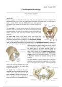
Pelvises of women and men differ in many ways. The pelvic inlet of women is circular compared to the
heart shaped pelvic inlet of men, with a less distinct promontory and broader alae. The pubic angle is
larger in women (80-85°) than men (50-60°). The ischial spines generally
don’t project as far medially into the pelvic cavity in women as they do in
men.
The pelvic inlet is a circular opening between the abdominal cavity and
the pelvic cavity. The promontory of the sacrum protrudes into it, forming
its posterior margin. On either side, the margin is formed by the alae of the
sacrum, sacro iliac joint, and linea terminalis, down to the pubic
symphysis.
The pelvic walls consist of the sacrum, coccyx, pelvic bones, two
ligaments and two muscles. The ligaments of the pelvic wall are the
sacrospinous and sacrotuberous ligaments. The sacrospinous ligament
is the smaller, triangular in shape, with its apex attached to the ischial
spine, and the base to the margins of sacrum and coccyx. The sacrotuberous ligament is triangular as
well, and superficial to the sacrospinous. Its base attaches from the posterior superior iliac spine, along
the dorsal aspect and lateral margin of the sacrum, onto the
dorsolateral surface of the coccyx. The apex is attached to
the medial margin of the ischial tuberosity. These two
ligaments stabilise the sacrum and convert the greater and
lesser sciatic notches into the greater sciatic foramen,
superior to the sacrospinous ligament, and lesser sciatic
foramen, inferior to the sacrospinous ligament. The muscles
of the pelvic wall are the obturator internus and the
piriformis.
The pelvic outlet is a diamond shaped aperture, anteriorly
defined by bone and posteriorly by ligaments. In the midline,
the boundary is the pubic symphysis, continuing with both
sides of the pubic arch. Terminal parts of the
urinary and GI tracts and the vagina pass
though the pelvic outlet.
The pelvic floor is formed by the pelvic
diaphragm and the perineal membrane and
muscles in the deep perineal pouch. The
pelvic diaphragm is formed by the levator ani
and the coccygeus muscles. It separates the
pelvic cavity above from the perineum below.
1
, lunedì 15 aprile 2019
The two levator ani muscles originate from each
side of the pelvic wall and join together in the midline.
They attach to the pelvic wall at the posterior aspect
of the pubis, the tendineous arch in the fascia of the
obturator internus, and the spine of the ischium. At
the midline the muscles blend together around the
anal aperture, and posterior to it the form the
anococcygeal ligament, which attaches to the
coccyx. Anteriorly, the levator ani muscles are
separated by a U-shaped gap called the urogenital
hiatus. This allows the passage of urethra and vagina through the pelvic diaphragm. The pelvic
diaphragm is nothing but the muscular portion of the pelvic floor, given by the levator ani and coccygeus,
shaped like a bows and attached superiorly to the pelvic walls. The two coccygeus muscles are
triangular and overlie the sacrospinous ligaments, completing the posterior part of the diaphragm.
The perineal membrane is a thick fascia attached to the bony
framework of the pubic arch. It is oriented in the horizontal plane
and has a free posterior margin Anteriorly there is a small gap
between the membrane and the inferior pubic ligament. The
perineal membrane is above related to a thin space called the
deep perineal pouch, which contains a layer of skeletal muscle
and various neuro-vascular elements. The perineal membrane
and adjacent pubic arch provide attachment for the roots of the
external genitalia. The urethra penetrates vertically through a
circular hiatus in the perineal membrane. In women the vagina
also passes through a hiatus in the perineal membrane, just posterior to
the urethral hiatus. Within the deep perineal pouch, a sheet of skeletal
muscle functions as a sphincter, mainly for the urethra. Anteriorly, a group
of muscle fibres surrounds the urethra, forming the external urethral
sphincter, moreover, two additional groups of fibres are present in women.
One forms the sphincter urethrovaginalis, around both vagina and urethra, the other forms the
compressor urethrae. Together with the external sphincter, they facilitate closing of the urethra. In both
men and women, a deep transverse perineal muscle on each side parallels the free margin on the
perineal membrane and joins with its partner at the midline. These muscles stabilise the position of the
perineal body.
The perineal body is a poorly defined connective tissue structure into which muscles of the pelvic floor
and perineum attach. It is positioned in the midline, along the posterior border of the perineal membrane,
to which it attaches.
2
The benefits of buying summaries with Stuvia:
Guaranteed quality through customer reviews
Stuvia customers have reviewed more than 700,000 summaries. This how you know that you are buying the best documents.
Quick and easy check-out
You can quickly pay through credit card or Stuvia-credit for the summaries. There is no membership needed.
Focus on what matters
Your fellow students write the study notes themselves, which is why the documents are always reliable and up-to-date. This ensures you quickly get to the core!
Frequently asked questions
What do I get when I buy this document?
You get a PDF, available immediately after your purchase. The purchased document is accessible anytime, anywhere and indefinitely through your profile.
Satisfaction guarantee: how does it work?
Our satisfaction guarantee ensures that you always find a study document that suits you well. You fill out a form, and our customer service team takes care of the rest.
Who am I buying these notes from?
Stuvia is a marketplace, so you are not buying this document from us, but from seller Greta96. Stuvia facilitates payment to the seller.
Will I be stuck with a subscription?
No, you only buy these notes for $2.99. You're not tied to anything after your purchase.