DN_Lectures:
Lecture 1: Neurophysiology & Neuroanatomy
-Brain Organisation = Processing + Storage relevant/NB info is processed simultaneously & stored in different parts of brain
- Regulation: brain controls thoughts/behaviours/emotions (in collaboration w somatic nervous system) + internal body
processes (in collaboration w automatic nervous system)
o E.g. PFC = inhibits impulses, send/receive info via gut-brain axis
o Hypothalamus = regulates stress in collaboration with pituitary & adrenal gland (HPA Axis)
- Integration: brain connects incoming info (sensory input) with already stored info from memory
- Prediction: brain uses prior knowledge to determine future outcomes does not process all info, only what is not
expected (aka prediction error) more efficient processing
- Lateralization: two hemispheres have partially different functions LH = understanding + producing language,
semantic memory RH: processing social + emotional stimuli, visuo-spatial orientation LH/RH control opposite
sides of body
- Cooperation & Competition: some brain parts compete and some cooperate competition = top-down (high>low) vs
bottom-up (low>high) high = frontal cortex / low = primary projection areas, brainstem, limbic system
- Small vs Large Brain Networks: brain is organized both structurally & functionally into complex networks
-The Nervous System = the Central Nervous System (CNS – brain + spinal cord) + Peripheral Nervous System (PNS – Somatic
Nervous System (SNS) & Automatic Nervous System (ANS)
Peripheral Nervous system:
- 1)Somatic Nervous System: Sensory & Motor neurons Sensory (afferent)
neurons carry messages from outside to CNS, specialized in transmitting
messages from eyes, ears, & sensory receptors to spinal cord & brain) Motor
(efferent) neurons carry messages from CNS to skeletal muscles – from brain &
spinal cord to muscles that control voluntary movements
- 2)Autonomic Nervous System: regulates internal environment of body (e.g. respiration/circulation) senses internal
functions, controls glands and involuntary muscles (heart, blood vessels, stomach) motivation, emotional behaviour
and stress response Sympathetic Nervous System + Parasympathetic Nervous System
o 2.1)Sympathetic Nervous System (SNS): physical activation fight-flight response
o 2.2)Parasympathetic Nervous System (PNS): slows body processes and maintains relaxation
Central Nervous System: Spinal Cord + Brain (cranial nerves, brainstem, forebrain)
- 1)Spinal Cord: motor & sensory nerves, organizes reflexes central pattern
generators (rhythmic movements: walk)
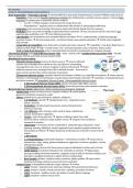
- 1)Brain: divided into 3 parts (forebrain, midbrain, hindbrain)
o 1.1) Hindbrain: vital life medulla (heart rate + respiration)/ pons
(sleep + arousal)
o Cerebellum: complex/rapid movements, precise timing (motor),
memory & learning
o 1.2) Midbrain: vision, hearing, motor control, sleep, wakefulness,
arousal, temperature regulation
o Tectum = input from eyes/ears Superior Colliculus (input from optic
nerve) & Inferior Colliculus (input auditory nerve) integrate info and directs
command to muscles
o Tegmentum= red nucleus (motor coordination), substantia nigra (dopamine, motor-
planning, learning, addiction), ventral tegmental area (complex synaptic network for
homeostasis & reflexes) largest dopamine-producing area involved in neural reward
system
o 1.3)Forebrain:
o 1.3.1)Hypothalamus: underneath thalamus = motivation & emotion sexual arousal,
temperature, sleep, eating, pleasure-displeasure, aggression, hormone regulation, contact
with pituitary gland)
o 1.3.2)Thalamus: receives info from sensory organs & distributes to other parts of brain
o Limbic System: coordinates behaviours for motivation & emotion hippocampus
(storing/recalling/retrieving memories), cingulate cortex (conflict in decision-making, error
detection), amygdala (response to salient stimuli, emotion/fear)
o Basal Ganglia: striatum, caudate nucleus, putamen, globus pallidus basal nuclei =
learning/controlling voluntary movements (not automatic)
- Cerebral Cortex: 2 hemispheres joined by corpus callosum 4 lobes:
o Lobe1: Frontal = planned motor, planning, thinking, WM, goal-directed behaviour
o Lobe2: Parietal = tactile, somatic sensory info, orienting, associative functions
o Lobe3: Temporal = auditory cortex, perception of social stimuli, language
o Lobe4: Occipital = primary visual cortex
, o Organisation of lobes: Lateral (outside)/medial (within/inside) Anterior (front)/Posterior (back) Dorsal
(top-back)/Ventral (down/front)
- Brain structure: cerebral cortex = higher cognitive functions separate brain regions
& their cell structure connect via white matter tracts to allow info exchange
different brain parts have different types of neurons
o gyri/sulci = grooves – sulci enlarge surface of cerebral cortex
o Neurons = 6 layer cell structure
o gray matter = cell bodies + dendrites of neurons reflect each part of body
(somatotopic map)
o white matter = fatty myelinated axons of neurons
o Role of different brain areas + how they cooperate global level (brain
networks), local level (brain area function), micro-level (neurophysiology –
neurons that fire)
o Projection Areas = receive info from senses or sends info directly to spinal
cord & muscles
o Association Areas = rest of the brain which does not have signals directly to
muscles/senses
- Brain Tissue: cells in the nervous system: 86 billion neurons + 1000 billion glial cells
o Glial cells: supporting cells, hold neurons in place, manufacture nutrients,
absorb toxins, guide neurons to position
o Neuron cells: cell body (soma) + neural branches (dendrites/axons) + axon terminal (synaptic terminal) +
myelin (white fatty tissue)
o Neural communication: dendrites receive signal from other neurons elicits electrical impulse in the axon
travels from dendrite through cell body to axon and exits via axon terminal
o # dendrites & spines determines the # synaptic connections
o Spines = surface of dendrites = small bulbs allowing more connections between other neurons
o Axon terminal: where chemical substance/neurotransmitter is released in the synaptic cleft/space
o Neural communication = electrical/chemical process: neurons generate electricity that create nerve impulses
& release chemicals to allow communication with muscles & glands
o Nerve impulses = action potentials chemicals = neurotransmitters
o Ions = electrically charged atoms outside axon = Sodium (NA+) & Chloride (CL-) ions inside axon =
proteins/anions (A-) + Potassium (K+) Uneven distribution of positive & negative ions inside vs outside cell =
state of polarization (difference = resting-potential) if stimulated, voltage shifts, leading to depolarization
causing an action potential (neuron fires – from negative to positive change)
o Action potential: channel opens and closes based on depolarization (e.g. chamber of opening/closing doors)
o Resting state = -70mV stimulation opens NA+ channels >> depolarization >> exceeds threshold >> action
potential >> NA+ will close & K+ will open (refractory period) >> neuron not able to discharge another impulse
o All-or-none law: action potentials only occur when it exceeds a threshold
o Graded potentials: many graded potentials together can elicit action potentials (depolarization which does not
exceed action potential threshold of -50mV (e.g. local anesthetics attach to sodium channels & block flow of
sodium ions into the neuron >> stop pain impulses).
o Neural Activity: 3 Basic Steps: (1) Resting Potential – distribution of positively & negatively charged ions
around axon (2) Action Potential – stimulation = ions flow into cell membrane channels = electrical charge
(3) Resting Potential – neuron restores ionic balance.
o Synaptic transmission: neurotransmitters carry messages across synaptic cleft 1. Synthesis >> 2. Storage (in
vesicles) >> 3. Release >> 4. Binding (neurotransmitter to receptor) >> 5. Deactivation (restored into axon)
o Excitatory neurotransmitters = cause post-synaptic neuron sodium channels to open >> depolarize
(graded/action potential)
o Inhibitory neurotransmitters = cause potassium to flow out of neuron or chloride into neuron >> polarization
(increase neurons negative potential – neuron will not fire – maintain state)
o Neurotransmitter types: (a) Acetylcholine (excitatory: muscle movement, memory) (b) Noradrenaline
(excitatory & inhibitory: learning, memory, wakefulness, eating) (c) Serotonin (excitatory & inhibitory: mood,
sleep, eating arousal) (d) Dopamine (excitatory: voluntary movement, emotion, learning, memory,
pleasure/pain) (e) GABA (inhibitory: motor) (f) Endorphin (inhibitory: pain) (g) Glutamate (excitatory)
o Dopamine projection pathways: ventral tegmental midbrain and axons can reach PFC & midbrain
o Serotonin projection pathway: transmitter substances widely distributed across brain, cerebellum, & spinal
cord
Lecture 2: Intro Brain & Plasticity
-Developmental neuropsychology: brain-behaviour relations of immature/developing brain in clinical practice ( 1980s)
-Developmental Cognitive Neuroscience: focus on normal development of cognitive functioning not same as developmental
neuropsychology (focus on abnormal)
, -Assumptions adult neuropsychology: adult brain = static/organised/less plastic 1:1 relation between structure & function
symptoms relate to neurological defect (functional localization)
-Assumptions developmental neuropsychology: immature brain = dynamic/spurts in brain related to changes in behavioural,
social and cognitive development symptoms and underlying neurological defect are not clearly related dependent on
timing of brain damage + social environment multiple factors: biological, cognitive, social-emotional, developmental &
environmental later outcome = difficult to predict address totality
-Anatomical Development:
(1) Zygote = fertilization 2 cells (undifferentiated)
(2) Morula = rapid cell division, cluster cells (undifferentiated)
(3) Blastocyst = differentiation inner cell mass = embryo / outer layer = placenta
14 days after fertilization inner cell mass of blastocyst develops 3 layers
- (3.1) Ectoderm = outer layer (Skin/CNS)
- (2) Mesoderm = middle (skeleton)
- (3) Endoderm = inner (intestines)
Neurulation/Neural Induction forms spinal cord neural plate created from ectoderm week 3-
4 = neural tube Closure Defects: causes = genetic/infection
- anencephaly = fatal closure defect of neural tube has subcortical structure but no
cortical structure)
- spina bifida = incomplete closing of backbone/spinal cord often combined with HYD
-Brain vesicles: telencephalon (cortex) diencephalon (thalamus/hypothalamus)
mesencephalon (midbrain) metencephalon (cerebellum/pons) Myelencephalon (medulla
oblongata)
-Development of Nervous System: different structures at different times (but overlapping) largely develops before birth
(1) spinal chord + brainstem (2) Amygdala, cerebellum & hippocampus (3) Thalamus & basal ganglia (4) Cerebral
cortex (posterior to anterior)
-Histological Development = cellular stages of almost all neurons (also overlapping stages) all same 5 stages occur for all
cortical areas (some faster than others) timing varies by brain region:
(1) Cell proliferation = create new neurons (2) Cell Migration = move to place of function (3) Cell differentiation & growth
= forming/developing (4) selective cell death & synaptic pruning (5) Myelinisation = efficiency
- (1) Proliferation (6-18 weeks): ventricles are hollow filled with CSF new neurons made inside proliferation zone (e.g.
ventricular zone & subventricular zone) progenitor cells (precursor cells): AKA Neuroblast and glioblast
o Problems (2-5 months) = causes: genetic/trauma
o neural defects = microencephaly (cell division stops) OR megalencephaly (overproduction cells) too small/
too large brain
o functional defects = motor/intellectual impairment, learning problems, epilepsy
- (2) Migration move to layer in cortex where cell will function passive migration = thalamus/brainstem active
migration = cortex – bypass older neurons
o Problems = causes: genetic/toxicity/infection/intrauterine damage
o neural defects = lissencephaly (smooth cortex, no sulci or gyri)
o Disorders of cell migration (1) Schizencephaly = abnormal cleft in cortex, cell layers not clearly defined (2)
Polymicrogyria = multiple small gyri, neurons in abnormal locations (3) Agenesis of Corpus Callosum =
absence (4) Dysplasia/heterotopia = abnormal cell layer structure/clump in wrong place
o Functional effects: epilepsy, motor/IQ/learning deficits/behavioural problems (severity varies w syndromes)
- (3) Differentiation: cells are at location and then form their function via differentiation different neurons have
different shapes & varieties (grow dendrites & axons) form synaptic connections (synaptogenesis)
- (4) Cell Death & Synapse Elimination = brain development overproduction + death of neurons & synapses
o Apoptosis = programmed cell death (brain & body)
o Pruning = synapse elimination (influenced by genes, experiences, hormones)
o Role of Experience: experience-expectant synapses (sensitive period) = visual system neurons need exposure
to visual input for synapses to survive and become functional (at certain time point) experience-dependent
plasticity (enriched environment) = not particular period but throughout development - need input from
environment (larger cortex & more connections)
o Neurons that fire together wire together = simultaneous activity of neurons strengthens connections
o Disorders in Synapse formation & Pruning: causes = genetic, toxic, stimulus/experience (?), problems during
migration/differentiation effect = none (synapses are flexible) OR abnormal development
Disorders of abnormal apoptosis: neurodegenerative disorders (ALS/ Alzheimer’s) – excessive apoptosis & ASD
(hypothesized) – slower in early childhood, excessive in older childhood & adolescence.
- (5) Myelinization = coating of axon fibers increases processing speed & efficient transmitting of info through brain
different rates in different parts of brain (quicker/earlier for motor/sensory but later/slower for cerebellar and
association areas)
o Disorders of myelinization: causes = genetic, toxic, trauma
The benefits of buying summaries with Stuvia:
Guaranteed quality through customer reviews
Stuvia customers have reviewed more than 700,000 summaries. This how you know that you are buying the best documents.
Quick and easy check-out
You can quickly pay through credit card or Stuvia-credit for the summaries. There is no membership needed.
Focus on what matters
Your fellow students write the study notes themselves, which is why the documents are always reliable and up-to-date. This ensures you quickly get to the core!
Frequently asked questions
What do I get when I buy this document?
You get a PDF, available immediately after your purchase. The purchased document is accessible anytime, anywhere and indefinitely through your profile.
Satisfaction guarantee: how does it work?
Our satisfaction guarantee ensures that you always find a study document that suits you well. You fill out a form, and our customer service team takes care of the rest.
Who am I buying these notes from?
Stuvia is a marketplace, so you are not buying this document from us, but from seller athenahaggiyannes. Stuvia facilitates payment to the seller.
Will I be stuck with a subscription?
No, you only buy these notes for $6.11. You're not tied to anything after your purchase.