ECG BASICS
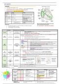
12-lead ECG:
• Praecordial (chest) leads (V1-V6)
• Limb leads (I, II, III, aVR, aVL, aVF) ® NORMALLY = aVR is ALWAYS NEGATIVE
• DO NOT ACCEPT ECG W/ ARTEFACTS OF BASELINE SHIFTS
• Progression of MI: LAD > RCA > Left circumflex
Location Leads Artery
Right atria V1, aVR
Inferior + AV II, III and aVF RCA è AV node + SA node
node
Antero-Septal V1-V4 LAD (MOST common)
Antero-lateral V4-5, I, aVL Left circumflex (LA + LV) =
major infarct
Lateral I, aVL +/- V5/6 Left circumflex ® marginal +
1st diagonal branch of LAD
Posterior • ST depression V1-3 Posterior desc artery (branch
• Dominant R wave V1-2 of RCA)
NB: left ventricular branch divides into the left
Intrinsic rate: anterior and left posterior branch
• SA node (60-100bpm) > AV node (40-60bpm) > Purkinje (20-40bpm) *RV = most anterior = most likely to be stabbed
• Slowest conduction = AV node
• R-R variability = SA node misfiring
Duration Mechanical event Pathology
atrial depol • ATRIAL HYPERTROPHY è Bifid “M” like = P mitrale ® LA enlargement due to MS
P wave < 0.12s • P Pulmonale è Peaked P wave > 2.5cm
(atrial arrhytmias)
• No P waves = AF / flutter / junctional rhythm
Shortened = Accessory pathway – WPW vs SVT (no P wave) è valsalvre, adenosine (cardiovert 1st if shock)
Prolonged = AVN dysfunction/block ® causes include:
P-R 0.12-0.2 s Conduction
• Inferior MI, electrolyte disturbance, increase vagal tone/athletes
interval (3-5 squares) (Heart blocks)
• AV blocking drugs (e.g. digoxin, amiodarone)
• Inflammation (IE)® autoimmune (SLE/SSc) ® infiltrative diseases (amyloidosis)
Narrow • SVT, atrial flutter, junctional escape
(supraventricular)
Broad • ectopic, LBBB or RBBB, wide complex tachycardias (VT, AF + BBB, torsades)
ventricular Very tall • S wave (V1) + tallest R wave (V5/6) ≥7 BIG squares ® LVH (HTN, AS, AR, MR, HOCM)
QRS depolarisation • Dominant R wave (V1) + Dominant S wave (V5/6) ® RVH (pulm HTN, MS, PE)
0.08-0.12s
complex (Ventricular strain
Short • effusion/tamponade, pneumothorax
or BBB)
Tall Q wave • established/previous full thickness MI
Normal • R wave progression (normally – most negative V1/dominant S ® most positive
V6/dominant R
• Dominant R wave in V1 (RVH, posterior MI, chronic lung disease)
• Shortened – Hypercalcemia, digoxin
< 0.45s
QT Lead II, V5, V6 • Prolonged (> 440ms) [Torsades de Pointes [polymorphic VT] -HypoCa, HypoK, HypoMg
interval (less than half
[Dictates HR] o Other Causes = TCAs, sotalol, amiodarone, low electrolytes, macrolides, anti-psychotics
of R-R)
o Corrected using ® anti-bacterials (e.g. quinolones (-acins) and macrolides (-mycins)
ST ischemia, • Elevation = STEMI (infarction or if in every lead ® pericarditis (concave), cardiac tamponade)
< 0.15s • Depression = NSTEMI, UA (ischeamia, posterior MI)
segment electrolyte issue
T-wave Inversion
• Normal in III, avR and V1 (Right leads)
0.1 – 0.25s • Pathological = ischemia, electrolyte
(usu. ventricular PE, RVH, LVH, BBB, digoxin Rx
T wave
1/3rd height of repolarisation • Biphasic
QRS) o Up ® down= ischeamia
o Down ® Up = HypoK
U wave 0.08s Normal or pathological (- hypokalaemia, hypothermia or with anti-arrhythmic)
J wave Osborne wave ® hypothermia, hyper Ca, SAH
Lead I AVF Normal Variant Pathology
• Children • RBBB • RVH (in PE, lung
RIGHT AXIS • Tall thin adults • Left posterior hemiblock disease, PHTN)
deviation • COPD (vertical heart) • Anterolateral MI (delayed • Na channel blockade
• Dextrocardia conduction on left side) • WPW syndrome
• LBBB
• Pregnancy, • LVH, ?HOCM
LEFT AXIS • Left anterior hemiblock
• obesity, ascites • VT
deviation (most common cause)
• Abdo distension, tumour • RV pacemaker,
• Inferior MI
• Lead transposition è can cause both RAD/LAD
EXTREME
• Hyperkalemia è can cause both RAD/LAD
AXIS • Emphysema
• Pacemaker
deviation
• VT
NORMAL ECG Ø HR > 100bpm (correct for age) Ø Inferior+lateral Q waves
FINDINGS IN PAEDS Ø Short PR/QT Ø RAD, inverted T waves in anterior leads (RV larger than Lv)
,