Notes of all the lectures given during the course Heart Failure and Therapy (minor Biomedical Topics in Healthcare). The document also contains many useful images that match the explanation of the course material. (vak voor o.a. gezondheidswetenschappen, gezondheid en leven, biomedische wetenschap...
LECTURES HEART FAILURE AND THERAPY
LECTURE 1: PSYCHIOLOGY OF THE CARDIOVASCULAR SYSTEM
EXCITATION-CONTRACTION COUPLING
FUNCTION OF THE HEART
- Pumping deoxygenated blood to the lungs
- Pumping oxygenated blood to all the organs in the body
- Together with blood vessels: providing adequate perfusion of all organs and tissues of the body
- Contraction and relaxation determine cardiac output
- The ability of the ventricle to relax is very important
o What is the main determinant of cardiac output: contraction or relaxation?
▪ It’s not only the force, but more importantly the amount of blood that can fill the
ventricle (by relaxation) → heart failure is more often a result of relaxation problems
- How can they be sustained => coordination of
contraction and relaxation of 2-3 billion CMs that must
contract or relax in order to have a heart rate
Excitation contraction coupling
- Contraction of the heart following electrical
stimulation of cardiomyocytes (link between
membrane depolarization and contraction) → this only
happens when all cells are stimulated electrically
AUTOMATION OF THE HEART
- The heart can beat independent of hormonal input (although it can get input which can affect the
heart rate, this isn’t necessary)
- Automation (the heart keeps on beating): spontaneous active, pacemaker cells
- If you remove your heart from your body and provide it with warmth it keeps beating → there is no
active component
- If you remove it from the body it will be 100 bpm → humans in general → the heart beat is reduced
(input out of the nervous system)
o Pacemaker cells produce an electrical signal → action potential (pacemaker cells have specific
action potentials)
Blue sodium flows in the cell (high out of the cell) (during rest: sodium channel is open: it is high outside and
low inside so sodium wants to flow in→ membrane potential increases: it is higher in the cell)(slow process,
1
,there are not many sodium channels)→ red: calcium flows in the cell (Treshold: voltage gated channel: it opens
at a certain value→ -40 or higher it opens and the calcium enters the cell because it is lower in the cell)→
yellow: potassium flows out of the cell
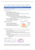
ACTION POTENTIAL CMS (CARDIOMYOCITES)
In SA node cells:
- At the threshold there’s a point of no return, so a full action potential is completed
- The electrical signal spreads through the atria and the ventricles
- Unstable resting potential → slowly becomes positive
→automation (reaches an action potential once in a while)
- Slow depolarisation
- Prepotential (pacemaker potential)
- Membrane potential is the difference in charge between the
inside and outside of the cell
In ventricular cells:
- Stabile resting potential: -85 mV → has to wait for other cells to produce an action potential
- Quick depolarisation
- Plateau
- Quick repolarisation
- Needs input from the AV node cell, otherwise nothing will happen →
is not on/off
- No automation, it only reacts when there is a signal
Basics for the resting membrane potential
- Membrane potential determined by concentration differences of ions and permeability to ions. It’s
largely determined by K+ gradient (see Nernst equation). The action potential is always negative. The
resting membrane potential is about -85 mV. Membrane potential (resting membrane potential and
action potential) is determined by:
o Sodium (Na+) and calcium (Ca2+) are always high outside cells → both want to go inside
o Potassium (K+) is always high inside cells → wants to go out
THE ION CHANNELS AND ACTION POTENTIAL OF VENTRICLE CELL
- The ion channels are sensitive for changes in ion concentrations
- The permeability to ions is determined by opening or closing ion channels: during rest only the
potassium channels are open
CMs only undergo an action potential when the neighbor undergoes an action potential → could be a
pacemaker cell or muscular system → when you isolate them it will not contract
Cardiomyocites can pass signals, but a bit slower than the conducting system does. Automation comes from
action potentials in cardiomyocytes. These cells are electrically coupled
There’s a signal. Neighbor cells have undergone an action potential, so there is an influx of ions. This leads to:
1. Opening of Sodium channels. They are voltage gated: they notice that there is a change in membrane
potential in the area and then they open. The positive charged sodium is attracted to the negatively
2
, charged cell. Sodium entery is very quick (fase 1) there is a quick rise → sodium channel opens when it
senses an change in memory potential → they open and close quickly
2. Calcium channels open: membrane potential (cell is negative)→ is a little slower (that’s why they have
a plateau)
3. Then potassium channels open: repolarization. The cell is not very negatively charged anymore →
potassium will leave the cell. Positively charged ions will go outside the cell, the membrane potential
goes down until it reaches its equilibrium again
➔ All these channels don’t need ATP → determined by concentration gradient
➔ The action potentials of ventricular cardiomyocytes are flat in the beginning, while pacemaker cells
have spontaneous depolarisation (prepotential) → they become a bit more positively charged
➔ Pacemaker cells are always slightly permeable for sodium and calcium
It is a long lasting action potential → stays on for a long time
and then goes off → this is because it can lead to blood filling
and pumping → contraction and relaxation → you don’t want
another signal to contract again → you want the signal to
pause before it can contract again→ because the signal is
given through all the CMs so it is in the whole ventricle
HEART RATE: PACEMAKER CELLS
Heart rate is determined by:
- Resting membrane potential of SA node cells
- Velocity of depolarization: slope of the prepotential
Heart rate can be changed by:
- Changing the lowest point/minimum point, which means changing the resting membrane potential →
you can change the resting membrane potential to a point that is closer to the threshold
- Changing the slope of the prepotential
- Your starting point can be regulated → if you start closer to your threshold, then you can reach
threshold earlier→ your heart rate increases (by exercising and stress) → that is because of the
hormone adrenalin (noradrenalin) → that opens the sodium channel → you start closer to your
threshold→ it starts earlier and becomes more steep
o Quicker depolarization and stepper pacemaker potential
o Less negative resting potential
- Decreasing heart rate: rest or digest: acetylcholine→ it opens your potassium channel→ it becomes
more negative → hyperpolarization
REFRACTORY PERIOD
Refractory period: period in which cells are inexcitable (Na+-channels are not reset)
- Absolute (stimulation doesn’t have an effect) and relative (only strong stimulation has an effect)
refractory periods
- Key to contraction/relaxation behaviour of cardiomyocytes → the function of the heart is dependent
on contraction and relaxation behaviour
- You don’t want the electrical signal to travel back to the cell where it started, so this is very important
EXCITATION CONTRACTION COUPLING
Contraction of the heart following electrical
stimulation of cardiomyocytes
Ca cycling in the heart: important to induce
contraction → we want the calcium rates to increase
in the cell (when you have excitation you have calcium
flowing into the cell, but this is not enough → calcium
induced calcium release): calcium entering in the cell
it binds to the RyR→ receptor of the SR: organelle
filled with calcium → then it will open and 10 times
more calcium will flow in to the cell→ to allow
relaxation the SR takes up the calcium immediately
after the release (this happens because of the SERCA)
4
, - C.I.C.R. = calcium induced calcium release Calcium is the signal that initiates contraction and
maintains contractions
- It’s an amplification system: when calcium comes into the cell from the ion channel, a whole host
of calcium is released from the sarcoplasmic reticulum (SR) → a lot of myofilaments can be
activated by thiscalcium
- To relax again, the cells need to get rid of the calcium
CA2+ AND CONTRACTION
Ca2+ binding to myofilament initiates contraction
Myosin is a thick filament, actin is a thin filament → they act with each other, but only if calcium is present
Myosin and actin want to bind to each other → they cannot connect because there is another protein that
blocks it → calcium will enter→ it binds to another protein → leads to change → pushes the other protein
away → actin filament is pulled by the myosin → you need atp to release myosin from actin because the
myosin wants to bind the actin really badly
- The binding site is normally locked by tropomyosin and the lock is controlled by the troponin complex
- Myosin and actin can only interact if calcium is binding to the troponin complex
- Once myosin heads and actin interact a power stroke will occur → pulls actin to one side
CONDUCTION SYSTEM
Pacemaker cell is on the right atrium → this is where the heart
beat starts → the pacemaker cells are located in the SA node
(sinoatrial)
AV ( atrioventricular) node: delay the signal from your atrium
to your ventricles: it can only enter your ventricles through the
AV node → slow conducting system (100ms delay)→ you don’t
want your ventricles to contract directly after your atria → it
conducts the electrical signal from the atria to the ventricles
- It is the only connection between the atria and
ventricles, they are isolated from each other
5
,SINGLE HEART BEAT (CELLULAR LEVEL)
- Electrical signal from neighboring cell (CM, SA node, conduction system)
- Action potential (Na+ influx → Ca2+ influx → K+ efflux)
- Ca2+ induced Ca2+ release
- Ca2+ binding to myofilaments
- Power stroke → cell shortening
- Ca2+-release from myofilaments
- Reuptake in SR → relaxation
If you increase the potassium levels outside the cell you will eliminate the concentration gradient. That way you
cannot depolarize. If you lower the sodium or calcium levels, you will also eliminate concentration gradient.
You therefore also take away the possibility to depolarize the heart.
THE CARDIAC CYCLE
Diastole: relaxation → filling the ventricle
Systole: contraction the ventricle
The heart injects blood into the aorta (high pressure) and presses blood to the right atrium to the ventricle
Ventricle pressure needs to be lower than the atrium pressure, but then it needs to be higher to pump the
blood into the aorta (aortic valves)
6
,PASSIVE FILLING: NO CONTRACTION
- Blood flows from your venous system from your right atria to the ventricles and
the aortic system to the left atria and ventricles
- AV valves: open
- Aortic/pulmonary valves: closed, otherwise blood flows back
- Both the atria and ventricles are relaxed, there’s no contraction
- In the ventricles the pressure is slightly lower than in the atria
ATRIAL CONTRACTION
- AV valves: open
- Aortic/pulmonary valves: closed
- Blood streams from the atria to the ventricles
- The atria are contracting (signal in SA node), while the ventricles are relaxed
- A small percentage of filling by pressure from the atria
ISOVOLUMETRIC CONTRACTION
- AV valves: closed→ pressure starts building, blood starts flowing back so the
valves close
- Aortic/pulmonary valves: closed because the pressure is not high enough to
eject but the pressure is higher than your atria
- The pressure in the ventricles increases quickly (above the pressure of the atria)
- A lot of pressure is built up (contraction of the ventricles), in order to open the
aortic and pulmonary valve (threshold needs to be reached)
Pressure starts building and building
EJECTION
- AV valves: closed
- Aortic/pulmonary valves: open
- Blood flows from the ventricles through the aorta and pulmonary artery
ISOVOLUMETRIC RELAXATION
- AV valves: closed
- Aortic/pulmonary valves: closed
- Both the atria and ventricles are relaxed
- After this it’ll go back to passive filling
7
, PRESSURE
VOLUME
End diastolic volume: relaxation: heart filles
End systolic volume: after you finish ejection: there is still blood remaining in the ventricle
Stroke volume: you pump certain amount out of your heart = end diastolic volume – end systolic volume
Ejection volume: stroke volume/EDV → used to be able to say something about the pump ability: heart failure
classification: how much of the blood that is in your heart can you pump out of your heart
Stroke volume= End diastolic volume – end systolic volume
Stuvia customers have reviewed more than 700,000 summaries. This how you know that you are buying the best documents.
Quick and easy check-out
You can quickly pay through credit card or Stuvia-credit for the summaries. There is no membership needed.
Focus on what matters
Your fellow students write the study notes themselves, which is why the documents are always reliable and up-to-date. This ensures you quickly get to the core!
Frequently asked questions
What do I get when I buy this document?
You get a PDF, available immediately after your purchase. The purchased document is accessible anytime, anywhere and indefinitely through your profile.
Satisfaction guarantee: how does it work?
Our satisfaction guarantee ensures that you always find a study document that suits you well. You fill out a form, and our customer service team takes care of the rest.
Who am I buying these notes from?
Stuvia is a marketplace, so you are not buying this document from us, but from seller Anouk152. Stuvia facilitates payment to the seller.
Will I be stuck with a subscription?
No, you only buy these notes for $5.38. You're not tied to anything after your purchase.