NSCA\\\'s Essentials of Personal Training 3rd edition
Summary
Samenvatting essentials of personal training 3e editie (hoofdstuk 1 t/m 4)
21 views 1 purchase
Course
NSCA\\\'s Essentials of Personal Training 3rd edition
Institution
Hogeschool Van Amsterdam (HvA)
Book
NSCA\'s Essentials of Personal Training
Hierbij een uitgebreide samenvatting van hoofdstuk 1 t/m 4 van NSCA essentials of personal training (3e editie), alle hoofdstukken zijn duidelijk omschreven en ik heb de minor afgerond met een gemiddelde van een 9. English: Here is an extensive summary of chapter 1 /tm 4 of NSCA essentials of perso...
Summary - Chapter 1-16 - NSCA's Essentials of Personal Training - Terms & Definitions
Samenvatting essentials of personal training 3e editie (Hoofdstuk 24 en 25)
Samenvatting essentials of personal training 3e editie (hoofdstuk 17 t/m 23)
All for this textbook (8)
Written for
Hogeschool van Amsterdam (HvA)
Minor personal trainer
NSCA's Essentials of Personal Training 3rd edition
All documents for this subject (7)
Seller
Follow
joepnorbart
Reviews received
Content preview
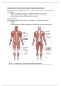
Chapter 1 structure and function of the muscular, nervous and skeletal systems.
Physical activity occurs through the combined and coordinated efforts of the muscular, nervous and
skeletal system.
- Nerves are responsible for initiating and modifying the actions of the muscles
- Muscle produce movement by generating forces to rotate bones around joints
- Skeletal rotation of bones provides movement of the body.
The muscular system
Force of a muscle called muscle contraction or muscle action. There are 3 types of muscles:
- Smooth
- Cardiac
- Skeletal: attaches to the bones, causing them to rotate (allows us to run, jump and lift etc)
,Gross anatomy (macrostructure) of skeletal muscle
Each skeletal muscle is surrounded by a layer of connective tissue referred to as epimysium. A muscle
is further divided into bundles of muscle fibers. A bundle of muscle fibers is called a fasciculus or
fascicle. Each fasciculus is surrounded by connective tissue called perimysium. Within a fasciculus,
each muscle fiber is surrounded and separated from adjacent fibers by a layer of connective tissue
referred to as endomysium. Together, these connective tissues help transmit the force of muscle
action to the bone via another connective tissue structure, the tendon.
Microsopic anatomy of skeletal muscle
Each muscle fiber is a cell, with many of the same structural components as other cells. Each muscle
fiber is surrounded by a plasma membrane, called a sarcolemma. A sarcolemma encloses the
contents of the cell, regulates the passage of materials such as glucose in and out of the cell and
receives and conducts stimuli in the form of electrical impulses or action potentials. Skeletal muscles
are multinucleated (possess more than one nucleus). A nuclei contain DNA from the cell and are
responsible for initiating the processes associated with adaptations to exercise. Within the boundary
of the sarcolemma, but outside the nuclei, is the cytoplasm, referred to as sarcoplasm in muscle. This
watery solution contains the cell’s energy source called adenosine triphosphate (ATP). Also
suspended within the sarcoplasm are organelles. These include mitochondria, which are the sites of
aerobic ATP production within the cell. Another important organelle is the sarcoplasmic reticulum,
this organelle stores calcium and regulated the muscle action process by altering the intracellular
calcium concentration. The sarcoplasmic reticulum releases calcium into the sarcoplasm of the cell
when action potential passes to the interior of the cell via structures called transverse tubulus (T-
Tubulus). T-tubulus are channels that form openings in the sarcolemma of the muscle cell.
, Myofibril
Each muscle cell contains columnar protein structures that run parallel to the length of the muscle
fiber, called myofibrils. Each myofibril is a bundle of myofilaments, which contain myosin (thick) and
actin (thin) filaments. Each myosin molecule consist of a head, neck and tail. The head is capable of
attaching to and pulling on the actin filament. ATP is used to perform the power stroke (important
step in the process of muscle activation). The neck structure connects the head to the tail. The
protein titin maintains the position of the myosin filament relative to actin.
Each actin filament is formed from individual globular, or G-actin. Each G-actin has a binding site for a
myosin head. There are two other protein structures in actin, tropomyosin and troponin. They
regulate the interaction of myosin and actin. Tropomyosin is a rod-like protein that spans the length
of seven G-actin proteins along the length of the actin filament. When the muscle is at rest,
tropomyosin lies of the myosin binding sites of actin. Each end of a tropomyosin filament is attached
to troponin. When bound to calcium, troponin causes the movement of tropomyosin away from the
myosin head to attach and pull on actin. Critical step in muscle activation. Protein nebulin acts to
ensure the actin filaments are the correct length.
Sarcomere
The sarcomere is the basic contractile unit of the muscle. It extends from one Z-line to an adjacent Z-
line. In the sarcomere there are actin and myosin filaments.
- Z-line: actin filaments anchored at one end of the Z-line. The extend inward to the centre of
the of the sarcomere.
- A-band: determined by the width of the myosin filaments. The A-band provides the dark
striation of skeletal muscle.
- H-zone: the area of the A-band that contains myosin, but not actin.
- M-line: in the middle of the H-zone is a dark line called the M-line. The M-line helps align
adjacent myosin filaments.
- I-band: the I band spans the distance between the ends of adjacent myosin filaments. Each I-
band lies partly in each of two sarcomeres.
The benefits of buying summaries with Stuvia:
Guaranteed quality through customer reviews
Stuvia customers have reviewed more than 700,000 summaries. This how you know that you are buying the best documents.
Quick and easy check-out
You can quickly pay through credit card or Stuvia-credit for the summaries. There is no membership needed.
Focus on what matters
Your fellow students write the study notes themselves, which is why the documents are always reliable and up-to-date. This ensures you quickly get to the core!
Frequently asked questions
What do I get when I buy this document?
You get a PDF, available immediately after your purchase. The purchased document is accessible anytime, anywhere and indefinitely through your profile.
Satisfaction guarantee: how does it work?
Our satisfaction guarantee ensures that you always find a study document that suits you well. You fill out a form, and our customer service team takes care of the rest.
Who am I buying these notes from?
Stuvia is a marketplace, so you are not buying this document from us, but from seller joepnorbart. Stuvia facilitates payment to the seller.
Will I be stuck with a subscription?
No, you only buy these notes for $8.62. You're not tied to anything after your purchase.