Diagnosis And Management Of Ocular Motility Disorders
This is a summary of the ocular motility conditions covered in the 3rd year of the Orthoptics degree at The University of Sheffield and particularly focuses on supranuclear conditions and nystagmus. These notes are perfect for Orthoptic students, Orthoptists, and any other professional/student inte...
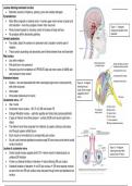
Lesions affecting brainstem function
• Brainstem consists of thalamus, pituitary, pons and medulla oblongata
Pyramidal tract
• Motor fibres originate in cerebral cortex → carries upper motor nerves to spinal cord
and brainstem – here they synapse to lower motor neurones Figure 1 – A diagram
• Fibres transmit signals for voluntary control of muscles of body and face showing the location of the
• No synapses within descending pathway brainstem
Cerebral peduncles
• Two stalks, attach the cerebrum to brainstem and is located in anterior part of
midbrain
• These contain ascending and descending nerve fibres between brain and brainstem
Red nucleus
• Lies within midbrain
• Pale pink due to iron presence
• Receives input from cerebellum of OPPOSITE side and motor cortex of SAME side
and involved in motor control
Brainstem lesions
• Nuclear – rare and associated with other neurological signs due to close proximity Figure 2 – A diagram
with other structures showing the sub
nuclei of the muscles
• Internuclear
supplied by the 3rd
• Infranuclear (below level of nuclei) nerve
Oculomotor nerve – 3rd
• Has 2 nuclei
• Oculomotor nerve nucleus – SR, IR, IO, MR and levator PS
• Erdinger-Westphal nucleus – sphincter pupillae and ciliary body (parasympathetic)
• 2 types of efferent nerve fibres present – somatic (EOM) and visceral (sphincter +
ciliary)
• The efferent nerve fibres originate from midbrain at superior colliculus and leaves
skull through superior orbital fissure
• Each muscle is innervated by its corresponding sub nucleus
• All sub nuclei innervate ipsilateral muscles except SR sub-nucleus and central caudal
nucleus (levators)
Figure 3 – A diagram
Lesions of oculomotor nerve
showing the nerve fibre
• Central caudal nucleus supplies both LPS → lesion results in bilateral ptosis c/s course of the 3rd nerve
unilateral SR limitation
• If there is a bilateral limitation of elevation → lesion affecting SR sub nucleus
• Unilateral limitation of elevation → not SR sub nucleus → SR nerve fascicle involved
as axons from one SR sub nucleus cross and pass through contra and ipsilateral sub
nucleus
,Lesions affecting brainstem function 2
Trochlear nerve – 4th
• Originates in midbrain and exits from posterior midbrain
• Smallest nerve by number of axons but has longest intracranial course
Figure 4 –
• Unable to distinguish between nuclear and fascicular lesions
A diagram
Abducens nerve – 6th showing
• Originates from the paramedian dorsal lower pons in floor of 4th ventricle lateral to the medial longitudinal the
fasciculus nucleus of
the 4th
• Nerve exits at junction of medulla and pons and courses over medial petrous apex towards cavernous
nerve
sinus
Causes of lesions of the 6th cranial nerve
• Brainstem syndrome
• Elevated ICP syndrome
• Petrous apex syndrome
• Cavernous sinus syndrome
• Orbital syndrome Figure 5 –
• Isolated 6th (microvascular) A diagram
Nuclear lesions of 6th nerve showing
• Horizontal gaze palsy where ipsilateral LR and contralateral MR are affected the
nucleus of
• 6th nucleus lies lateral to medial longitudinal fasciculus → some neurones project to MLF and cross over to the 6th
contralateral side and innervate contralateral MR sub nucleus nerve
Fascicular lesions of 6th nerve – ipsilateral LR palsy
Brainstem syndromes
• Caused by lesions such as infarction, haemorrhage, tumour, demyelination, trauma Figure 6 – A
diagram showing
• Causes multiple cranial nerve involvement
the projection of
• Weber’s – midbrain stroke syndrome, 3rd nerve fascicles and cerebral peduncles affected fibres of MLF to 6th
o Ipsilateral 3rd NP and contralateral hemiparesis nerve nuclei,
• Benedikt’s – paramedian midbrain syndrome, 3rd nerve fascicles, red nucleus and cerebral peduncle causing a
horizontal gaze
affected palsy
o Ipsilateral 3rd NP, contralateral hemiparesis, contralateral ataxia with hyperkinesis/tremor
• Foville’s – abducens nucleus, anterior pons and pyramidal tracts affected
o Ipsilateral nuclear 6th NP, ipsilateral horizontal gaze palsy, ipsilateral facial palsy, contralat.
hemiparesis
• Millard-Gubler – base of pons, antero-medially affecting 6th and 7th nerve fascicles and pyramidal tracts Figure 7 –
A diagram
o Ipsilateral 6th nerve palsy, ipsilateral facial nerve palsy, contralateral hemiplegia
showing
Collier’s sign Collier’s
• Unilateral or bilateral lid retraction due to midbrain lesions – characteristic feature of dorsal sign
midbrain/Parinaud’s syndrome
• Also upward gaze palsy, convergence retraction nystagmus, bilateral lid retraction and light-near
dissociation
,Lesions affecting brainstem function 3
Divergence paralysis
• Poorly understood – theories suggest there to be a divergence centre around the 6th Figure 8 – A diagram
showing the 3rd nerve
nerve nucleus and that it is due to a lesion of the cerebellum or Arnold-Chiari
fascicles, red nucleus
malformation and cerebral
• Signs are ET, homonymous diplopia, normal OM but absent negative fusion amplitude peduncles being
• Aetiology is raised ICP, MS, encephalitis, trauma, Miller-Fisher syndrome affected in Benedikt’s
• Differential diagnosis – 6th NP, concomitant ET, convergence spasm
• Treatment – may resolve → observe, occlusion or BO prisms or LR resections as last
resort
Diseases affecting brainstem and OM function
• Parkinson’s – degenerative, insufficient dopamine production – causes are idiopathic,
viral, inherited and drug-induced Figure 9 – A diagram
o Rigidity, tremors are most recognised characteristics – no cure showing the 3rd nerve
o Ocular features – lim. elevation, dep. Affected later, hypometric saccades, CI, fascicles and cerebral
nystagmus, reduced control of phoria, impaired smooth pursuit, blepharospasm, peduncles being
affected in Weber’s
lid lag
• Huntington’s – hereditary, substantia nigra may be involved in brain stem
o Characteristics are loss of mobility, speech and swallowing difficulty
o Ocular features – difficulty initiating saccades, slow saccades, impaired smooth
pursuit
• Wernicke’s encephalopathy – caused by thiamine deficiency, common in alcoholics
and GI disorders Figure 10 – A diagram
o Characteristics are gait ataxia, impaired short term memory – where it is not showing the 6th
nucleus, anterior pons
treated timely, it may progress to Korkasoff’s syndrome which is irreversible
and pyramidal tracts
o Ocular features – abduction weakness, gaze evoked nystagmus, INO, vertical affected in Foville’s
nystagmus, horizontal and vertical gaze palsies, complete ophthalmoplegia
• Whipple’s – Tropheryma whippelii bacteria → weight loss, diarrhoea, GI bleeding, joint
pain etc.
o Treated with antibiotics, bacteria may remain in CSF and fatal if untreated
o Ocular features – reduced vertical saccades, vertical and horizontal gaze palsies
and pendular oscillations
• Arnold-Chiari malformation – anomaly where cerebellar tonsils are displaced Figure 11 – A diagram
downwards towards foramen magnum and may herniate – congenital and acquired showing the 6th and 7th
fascicles, pons and
o Characteristics are head ache, neck pain, tinnitus, facial pain, muscle weakness, pyramidal tracts being
swallowing difficulty, sleep apnoea, impaired coordination, rapid heartbeat, affected in Millard-
dizziness etc. Gubler
o Ocular features – nystagmus especially downbeat, impaired pursuit, impaired
OKN, concomitant ET, divergence paralysis, skew deviation, INO
, Localisation of lesions and life-threatening emergencies 1
• Cerebrum consists of complex neural pathways which process
visual information to coordinate eye movements
• Brainstem is the main cerebral structure containing ocular motor
nuclei and gaze centres
Localisation of function
• Certain areas of the brain are responsible for certain function
• Motor cortex – movement and somatosensory inputs
• Visual cortex – visual information processing
• Broca’s and Wernicke’s area – speech production and
comprehension
Hemispheric lateralisation
• Each hemisphere is specialised to perform certain functions – R
for spatial perception and memory, L for language
Figure 1 – A diagram showing the idea
• Corpus callosum, connects pathways between each hemisphere of hemispheric lateralization – R with
• R side able to compensate for L but L not able to for R → L sided blue and L with red
visual inattention
Function of eye movements
• Eye movements are organised in a hierarchy that descends
down from the most superior cerebral regions to the eyes itself
Figure 2 – A diagram showing the hierarchical
• Pathway is supranuclear centres → brainstem → Infranuclear organization of eye movements, specifically,
pathways → EOM smooth pursuit
Control of eye movements
• Smooth pursuit, saccades, VOR, OKN and vergence
• Pupils and lids
Localisation of OM defects
• Some signs of ocular motility disturbances can localise a lesion
→ indicate affected area Figure 3 – A
• Some signs are non-localising signs and some are false- diagram showing all
localising signs the possible 6th CN
lesions that could
False localising signs – 6th CN
occur and their
• Unilateral or bilateral locations
• Often occurs in context of raised ICP → SOL, idiopathic
intracranial hypertension, cerebral venous thrombosis
• Mechanism → debated, though could be due to long intracranial
course, compression against ridge of petrous temporal bone or
the effect of backwards brainstem displacement
The benefits of buying summaries with Stuvia:
Guaranteed quality through customer reviews
Stuvia customers have reviewed more than 700,000 summaries. This how you know that you are buying the best documents.
Quick and easy check-out
You can quickly pay through credit card or Stuvia-credit for the summaries. There is no membership needed.
Focus on what matters
Your fellow students write the study notes themselves, which is why the documents are always reliable and up-to-date. This ensures you quickly get to the core!
Frequently asked questions
What do I get when I buy this document?
You get a PDF, available immediately after your purchase. The purchased document is accessible anytime, anywhere and indefinitely through your profile.
Satisfaction guarantee: how does it work?
Our satisfaction guarantee ensures that you always find a study document that suits you well. You fill out a form, and our customer service team takes care of the rest.
Who am I buying these notes from?
Stuvia is a marketplace, so you are not buying this document from us, but from seller q-abdulqadir. Stuvia facilitates payment to the seller.
Will I be stuck with a subscription?
No, you only buy these notes for $28.28. You're not tied to anything after your purchase.