Test Bank - Biopsychology, 11th Edition (Pinel, 2021) Chapter 1-18 | All Chapters
Test Bank for Biopsychology, Global Edition 11th Edition by John Pinel, Steven Barnes, All Chapters |Complete Guide A+
Test Bank - for Biopsychology, 11th Edition by John Pinel, All Chapters | Complete Guide A+
All for this textbook (18)
Written for
University of Greenwich
Unknown
Psychology
All documents for this subject (15)
Seller
Follow
soniakhan
Content preview
Chapter 3 – anatomy of the nervous system
The body has two nervous systems:
The peripheral nervous system and the central nervous system. The CNS consists of the brain and
the spinal cord, whereas the PNS consists of the body parts outside of these structures, so the limbs
and torso.
The peripheral nervous system consists of the somatic nervous system and the autonomic nervous
system.
The somatic nervous system controls responses to external stimuli, so when someone touches you
or blows wind in your eyes. Afferent neurons (sensory neurons) carry sensory signals from the skin,
muscles, skeletal muscle and glands to the central nervous system, which are then processed and
sent as efferent neurons (motor neurons) back to the skin or muscle (where the stimuli were
detected) to be picked up and responded to, usually involuntary.
The autonomic nervous system controls internal environments. It causes afferent neurons to move
from internal organs to the CNS, where efferent neurons then send signals from the CNS back to the
internal organs. The autonomic nervous system comprises of two smaller systems: the sympathetic
nervous system and the parasympathetic nervous system. The sympathetic nerves project from the
CNS in the lumbar (lower back) and the thoracic (chest/upper/middle back) regions of the body. The
parasympathetic nerves project from the brain and (sacral) lower back.
Sympathetic Parasympathetic
Mobilises energy resources and instigates fight Conserves energy and relaxes the body
or flight
Arousal Equilibrium
Triggers aroused response in organ, like Maintains regular response in organ, like rest
elevated heart rate and BP heart rate
Salivary gland: decreases secretion Salivary glands: increase secretion, easy to eat
Blood vessels constrict which then increases Blood vessels dilate, making blood flow easier
BP
Penis ejaculates Penis gets erect
Iris dilates the pupil Iris constricts
Sweat glands are stimulated Sweat glands are not stimulated
Tear glands are not stimulated Tear glands can produce moisture
Arrector pili muscles cause hairs to stand up No goosebumps
(Goosebumps)
Lungs inhibit mucus secretion and dilate Lungs stimulate mucus production and
bronchioles, allowing one to take more breaths bronchioles constrict
Cranial nerves: They project from the brain instead of the spinal cord, unlike every other PNS nerve.
They involve mostly sensory nerves, like olfaction and optic nerves, but also have motor neurons
present. Vagus nerves are the longest, with motor and sensory neurons going to and from the gut.
Olfactory (smell), optic (visual messages), oculomotor (coordinate eye movement), trochlear
(controls eye movement), abducens (lateral eye movement, to allow abduction), trigeminal (skin,
sinuses and mucus membranes), facial, glossopharyngeal (tongue and throat), vestibulocochlear
branch (balance), vagus (gut), spinal accessory (neck) and hypoglossal (base of brain to the tongue).
, If there is any notable damage to regions connected to the vagus nerve, then we can conduct scans
and understand where underlying tumours or medical conditions may be based depending on what
has been impaired, so losing senses of smell means olfaction is affected.
Meninges
Three protective membranes that, along with bones, encase the internal organs. The outer meninx
is dura mater (tough membrane; skull region, supplies large venous channels that supply blood from
the brain to the heart), the middle consists of arachnoid membrane (spider like membrane,
surrounds CSF source SAS) and beneath this, is a small space called subarachnoid space (protects
brain from sudden injury), containing spinal cerebrospinal fluid and large blood vessels, which carry
nutrients and line the brain and spinal cord. The innermost meninx is the pia mater, which is on the
surface of the CNS. It helps pass blood vessels through the brain.
Ventricles and cerebrospinal fluid
Cerebrospinal fluid protects the brain and spinal cord, located in the central canal, subarachnoid
space and central ventricles. The subarachnoid space is in the middle of the pia mater and dura
mater, the central canal runs across the whole spinal cord and the central ventricles are 4 chambers
of the brain. When cerebrospinal fluid leaks or is drained, people experience piercing headaches due
to susceptibility to external stimuli and lacking protection to the brain.
Cerebrospinal fluid is produced by choroid plexuses, which are small blood vessels that protrude into
the ventricle chambers through the pia mater. Any excess cerebrospinal fluid is absorbed from the
subarachnoid space into the Dural sinuses, which drains fluids, causing it to flow into the jugular
vein, which collects cranial blood, pumping it to the heart. If a tumour located near the narrow
channels connecting the ventricles, then this can cause a blockage in CSF flow, which leads to
hydrocephalus.
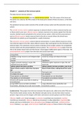
Scalp
Skull
Dura mater
Arachnoid
meninx
Subarachnoid
space
Sinuses
Cortex
Central canal is near the brain stem and
carried CSF to the spinal cord, 4th ventricle is
near the pons, cerebral aqueduct is in the
midbrain, the 3rd ventricle is near the
Artery hypothalamus and below the corpus
Pia collosum, and the lateral ventricles are in the
Mater cerebrum. They all hold cerebrospinal fluid,
which can get blocked in the CA.
The benefits of buying summaries with Stuvia:
Guaranteed quality through customer reviews
Stuvia customers have reviewed more than 700,000 summaries. This how you know that you are buying the best documents.
Quick and easy check-out
You can quickly pay through credit card or Stuvia-credit for the summaries. There is no membership needed.
Focus on what matters
Your fellow students write the study notes themselves, which is why the documents are always reliable and up-to-date. This ensures you quickly get to the core!
Frequently asked questions
What do I get when I buy this document?
You get a PDF, available immediately after your purchase. The purchased document is accessible anytime, anywhere and indefinitely through your profile.
Satisfaction guarantee: how does it work?
Our satisfaction guarantee ensures that you always find a study document that suits you well. You fill out a form, and our customer service team takes care of the rest.
Who am I buying these notes from?
Stuvia is a marketplace, so you are not buying this document from us, but from seller soniakhan. Stuvia facilitates payment to the seller.
Will I be stuck with a subscription?
No, you only buy these notes for $8.76. You're not tied to anything after your purchase.