Unit 3 - Anatomy and Physiology for Health and Social Care
Institution
PEARSON (PEARSON)
Around 90 pages of notes for the Anatomy and Physiology Exam that will help with revision and making sure that you are covering all of the specifications.
Unit 3 - Anatomy and Physiology for Health and Social Care
All documents for this subject (32)
1
review
By: keiratodd • 10 months ago
Seller
Follow
maisiesmith1
Reviews received
Content preview
Anatomy and physiology for health and social care
A – the structure and organisation of the human body
A1- how cells work
Cells
• There are about 50 trillion cells in the body.
• Your life began as a clump of eight stem cells.
• Each stem cell had the potential to develop into any different cell that makes up the
body.
• Over time they divide and differentiate into specialist cell types such as: nerve,
bones and muscle cells.
• Each will have its own function
How cells work:
• Cells carry out chemical reactions and processes that make up life itself
• They are microscopic and the basic unit of living material
• Cells rarely exist in isolation
Cells → tissues → organs → organs or body systems
The largest cell in the human body is the female ovum
Looking at cells
• Ordinary light microscopes are quite good for
viewing tissues and organs
• Electron microscopes are needed to see the detail
of cell contents
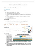
Cell membrane
• The cell membrane
is a double layer of
lipids and proteins
that surround a cell.
The lipids are small
fatty molecules in
two layers (bilayer)
with larger protein molecules inserted at intervals partly or completely through the
bilayer.
• The lipid molecules are phospholipids, the phosphate head is water soluble and the
lipid chains are insoluble. This is why they align themselves with chains facing away
from one another.
, • The fluid surrounding cells (tissue fluid) and cytoplasm are both watery
environments next to the phosphate heads.
• Protein molecules form channels for substances to pass too and from the cell.
• The protein molecules act as identify markers or reception sites for other molecules
such as hormones that are important to that cell
• This structure is often termed the ‘fluid mosaic model’ of the cell membrane
Cytoplasm
Cytoplasm is a semi-solid fluid, a bit like a gel, capable of
flowing slowly. Many chemical reactions are carried out in the
cytoplasm. The collective terms for these reactions is
metabolism. Complex sugars such as glycogen and melanin,
the dark pigment responsible for skin and hair colour are found
in cytoplasm.
Nucleus
• This is usually the largest structure inside a
cell. When viewed under a microscope it
stands out as a dark shape as it takes up
dyes and stains very easily.
• Most cells have a single, central, spherical
nucleus, but there are many variations.
• Some muscle cells have many nuclei and
are called ‘multinucleate’
• Red blood cells and platelets do not have a nucleus and are said to be ‘anucleate’
• White blood cells have a distinct, lobbed nuclei.
o Apart from red blood cells, which have a limited
life span and cannot reproduce most cells
separated from their nucleus will die.
o The nuclear membrane has a structure similar to
that of the cell membrane, but with gaps and
pores through which proteins and nucleic acid can
pass.
,The cell is said to go through cycles of division (Mitosis), replication (synthesis) and resting
(interphase).
When the cell is not dividing it is resting – interphase, and the nuclear material appears like
a dark, tangled mass and is called the chromatin network.
A smaller, darker sphere is often visible, the nucleolus. This is a source of ribonucleic acid
(RNA), one of the nucleic acids.
• There are 23 pairs of chromosomes in a human cell, containing specific
sequences of deoxyribonucleic acid (DNA) another nucleic acid.
• DNA is responsible for all our inherited characteristics, such as hair and eye
colour. The sequences of DNA are our genes.
• The nucleus controls nearly all the activities of the cell and has been likened to
the architectural drawing or blueprint front which the cell operates
, The structure and function of cells
You will learn about a typical human cell (animal cell), the details of the cell are known as
the ultrastructure of the cell. (Animal cells are also known as Eukaryotic cells,
these are cells that contain a nucleus and organelles, and are enclosed by a plasma
membrane)
A typical cell only exists for study purposes, it is not specialised. When studying actual cells
in the body you adapt this knowledge to the specific cell that is being considered.
For example, a mature red blood cell does not have a nucleus so if you were describing a red
blood cells ultra-structure you would not include the nucleus
Organ – a collection of tissues joined together to carry out a particular function
Organelle – a tiny body inside a cell, which carries out its own functions
Centrioles
• Every cell in the body has two small
organelles called centrioles. Centrioles play
a part in cell division and are usually found
near the cell nucleus lying at right angles to
each other.
• They cannot been seen unless the cell is
dividing when they may be seen through a
microscope, as two black dots.
• They are made of protein strands called
microtubules which move to opposite ends
of the cells at the start of cell division. Here
they make even more tubules which are known as the miotic spindle
• These threads connect to chromosomes to give the new cells formed the correct
amount of DNA.
Mitochondria
• Every cell in the body has at least 1000 rod-
shaped or spherical bodies, known as
mitochondria, which are concerned with energy
release. Very energy –active cells (like muscle
and liver cells) will have many more.
• Each mitochondrion has a double-layered
membrane, like the cell membrane but the
inner layer is folded at intervals, producing a
series of shelves or ridges, known as cristae.
• The enzymes responsible for end stages of glucose oxidation (or cell respiration) are
located on the cristae.
The benefits of buying summaries with Stuvia:
Guaranteed quality through customer reviews
Stuvia customers have reviewed more than 700,000 summaries. This how you know that you are buying the best documents.
Quick and easy check-out
You can quickly pay through credit card or Stuvia-credit for the summaries. There is no membership needed.
Focus on what matters
Your fellow students write the study notes themselves, which is why the documents are always reliable and up-to-date. This ensures you quickly get to the core!
Frequently asked questions
What do I get when I buy this document?
You get a PDF, available immediately after your purchase. The purchased document is accessible anytime, anywhere and indefinitely through your profile.
Satisfaction guarantee: how does it work?
Our satisfaction guarantee ensures that you always find a study document that suits you well. You fill out a form, and our customer service team takes care of the rest.
Who am I buying these notes from?
Stuvia is a marketplace, so you are not buying this document from us, but from seller maisiesmith1. Stuvia facilitates payment to the seller.
Will I be stuck with a subscription?
No, you only buy these notes for $14.37. You're not tied to anything after your purchase.