NEUR0014 Neural Basis of Motivation and Learning (NEUR0014)
Institution
University College London (UCL)
Notes and additional reading for the module including textbook notes, assigned reading, and extended reading. Evidence and research papers allocated in tables (note, not all research papers are described, but titles and authors are provided as references). Broken down into chapters and subchapters....
NEUR0014 Neural Basis of Motivation and Learning (NEUR0014)
All documents for this subject (1)
3
reviews
By: inajardevall • 5 months ago
By: brdpkd2222 • 1 year ago
By: merrywang2001 • 1 year ago
Seller
Follow
denh11323
Reviews received
Content preview
NEUR0014
NEURAL BASIS OF MOTIVATION AND LEARNING
The Limbic System
EMOTION
- Emotions are the result of the brain receiving feedback from the body
- Different emotions correspond to different sets of bodily changes\
JAMES-LANGE THEORY OF EMOTION
Emotions occur as a result of physiological reactions to events (you don’t run from the lion because you are afraid,
you are afraid because you run from the lion)
Fear is your body responding to its internal signals
Criticisms of James-Lange Theory
- Bodily changes too slow and non-specific
- Bodily changes not required for emotional behaviors
- Work by Philip Bard showed that disconnecting the cortex from the rest of the brain leaves emotional
displays intact
CANNON-BARD THEORY OF EMOTION
Emotional responses are not caused by conscious emotional experiences
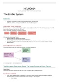
THE HISTORICAL EMOTIONAL BRAIN: THE LIMBIC SYSTEM OR PAPEZ CIRCUIT
Papez Circuit
Papez was the first to introduce the idea that limbic structures might mediate emotions
KLUVER-BUCY SYNDROME
- 1937: while studying effects of mescaline on the brain, Kluver and busy found that bilateral temporal
lobectomy rhesus monkeys displayed: → due to damage to the amygdala
o Visual defects
, o Oral tendencies
o Changes in emotional behaviour
HOW THE LIMBIC LOBE BECAME THE LIMBIC SYSTEM
1. Broca’s ‘lobe lymbique’
2. Papez’s emotional brain circuit
3. MacLean’s ‘emotional keyboard’ (1949)
a. Drawing from Papez’s theory, Kluver and Bucy’s experiments and clinical data, introduced the idea
that emotions are mediated by the visceral brain (1949) which he later (1952) referred to as ‘limbic
system’
The Truine Brain (Paul MacLean, 1970)
Limits of the Limbic System Concept
- Through the years the limbic system has expanded to encompass many more brain regions than those first
mentioned by MacLean and Papez, for some this means that the limbic system concept has lost specificity.
- The evolutionary account proposed by MacLean has been discredited. The brain is not made up of different
layers some more primitive than others.
- The limbic brain is not exclusively connected to the hypothalamus. The hypothalamus is connected pretty
much with the whole brain (more on this later).
- Some regions of the limbic system appear to be more involved in memory (and spatial navigation) rather
than emotional processing (anterior thalamus, mammillary bodies, hippocampus)
Basic Anatomical Components of Limbic System:
- Parahippocampal gyrus
- Olfactory cortex
- Hippocampus
- Cingulate gyrus
- Caudal orbital and medial prefrontal cortex
- Amygdaloid nuclear complex
- Antero-ventral insula
MODERN NEUROSCIENCE OF THE LIMBIC SYSTEM AND THE EMOTIONAL BRAIN
Korsakoff’s Syndrome
People who abuse alcohol suffer from atrophy of mamillary bodies and therefore memory loss
Bilateral temporal lobectomy – Scoville and Milner (1957)
MOTIVATION – BASIC CONCEPTS AND BRAIN MECHANISMS
,Hypothalamus: Behaviour Control Centre
GROSS ANATOMY AND FUNCTION
Basic Functions
- Regulation of physical state (homeostasis/adaptive)
- Control of several survival critical behaviours
- Learning, memory, attention, etc.
Hypothalamus is comprised of a collection of nuclei with relatively distinct functions
Inputs
Nucleus solitarius: provides input from ANS, involved in mediating autonomic feedback responses from
hypothalamus
- Contains baroreceptors to regulate blood pressure
Outputs
ENDOCRINE CONTROL
- The posterior pituitary (neurohypophysis) is comprised of secretory nerve terminals, the bodies of which
reside in hypothalamic nuclei
o Peptide hormones are released into the portal vessel system
- The anterior pituitary (adenohypophysis) is comprised of non-neuronal secretory cells
o Neurotransmitters and releasing factors are released into the portal vessels upstream of the
adenohypophysis which regulate hormonal release.
- Different hypothalamic nuclei regulate the release of different specific hormones (Kovacs & Ojeda, 2012)
o TRH: released by paraventricular nucleus → viral stimulating hormone on pituitary → increase
metabolism
o GHRH: released from nuclei (arcuate nucleus) → triggers release of growth hormones
, o GnRH: released from cluster of neurons around median eminence → developed in the vasal
epithelium and migrate into the olfactory bulb during development + settle in hypothalamus →
during puberty, become active and stimulate release of puberty and reproductive hormones
INGESTIVE BEHAVIOUR OF HYPOTHALAMUS
TWO-CENTRE HYPOTHESIS
1. Lateral hypothalamus: feeding center
a. Lesions promote satiety
b. Stimulation induces feeding
c. Hunger increases activity
d. Feeding centre
2. Ventromedial hypothalamus: satiety centre
a. Lesions cause hyperphagia
b. Stimulation inhibits feeding
c. Hunger reduces activity
d. Satiety centre
LATERAL HYPOTHALAMUS
Classic experiments on LHA function
1. Lesion studies in 1940’s-1980s showed that electrolytic lesions of LHS suppressed feeding and drinking while
lesioning of nearby VMH promotes feeding and body weight gain
2. Chemical lesions destroying catecholaminergic fibres containing either NA or DA demonstrated that these
fibres of passage contained within median forebrain bundle are important for feeding and drinking
3. Chemical lesions ablating LHA somata but spare passing fibres suppress feeding and drinking
4. Electrical stimulation of LHA in rodents showed that gross electrical activation of this region produced
voracious feeding behaviour (Delgado and Anand, 1953) and reinforced lever-pressing behaviour to gain
additional stimulation (Olds and Milner, 1954)
a. When animals are given control of a stimulating electrode in the base of this region, they choose to
stimulate it perpetually → overeating and strong sensation of reward → eat themselves to death
5. Intra-LHA injection of NT agonists or antagonists demonstrated that glutamate receptor activation induces
feeding while GABA agonist can suppress it
Conclusion; LHA and associated brain regions are critical for feeding and other drive-like effects and also for
reinforcement processes
4 neuronal subtypes linked to appetite behaviour (Stuber and Wise 2016)
1. Orexin → promote feeding and arousal
2. MCH → promote feeding and sleep
3. VGAT → GABAergic neurons that promote feeding
4. VGLUT → inhibit feeding (antagonistic effect)
Evidence of neuronal subtypes linked to appetite behaviour
1. Optogenetic Evidence
a. Jennings et al., 2013: Photostimulation of GABAergic projections from the BNST to VGLUT neurones
in the lateral hypothalamus induces eating in a well-fed and satiated mouse
Aim - BNST is a key integrator of motivational states through interaction with
LH
- BNST is comprised mostly of GABAerfic cells and consumption of food
activates BNST neurons (Angeles-Castellanos et al., 2007)
- Considered BNST and inhibitory projections to LH as important
candidate of regulated feeding
Method - ChR2-eYFP into BNST of Vgat-ires-Cre mice and optical fibres
positioned above LH for photostimulation of the Vgat BNST → LH
, projection fibres
Results - Optogenetic activation of the pathway produced voracious feeding
behaviour in well-fed mice
- Mice demonstrated place preference for photostimulation-paired
chamber
- Food deprivation augmented and satiety significantly attenuated self-
stimulation
- Photoactivation of the projections DID NOT elicit feeding behaviour
- Optogenetic stimulation of VGlut2 expressing LHA neurons has
opposite effect, reducing feeding in hungry mice and producing
aversion to locations where stimulation of these cells occurs
Conclusion Inhibitory inputs from the BNST specifically innervate and suppress LH
glutamatergic neurons to promote feeding ==? Therapeutic intervention within
these circuits to treat eating disorders and obesity
Critical
thinking
b. Jennings et al., 2015: Direct optogenetic activation of VGat expressing LHA neuron produces
voracious feeding and optical self-stimulation behaviour – reminiscent of electrical stimulation of
LHA
Aim Based on earlier study that optogenetic modulation of LH glutamatergic
neurons influences feeding and motivated behavioural responding
- Now examined if molecularly defined LH neurons that express Vgat
and synthesis and release GABA selectively promote and encode
appetitive and consummatory behaviours
Method -
Results - Show that selective optogenetic stimulation of LH GABAergic Vgat-
expressing neurons enhances appetitive and consummatory
behaviours and genetic ablation reduces these phenotypes
- Targeted LH subpopulation is distinct from cells containing MCH and
orexin
- LH GABAergic neurons preferentially encode aspects of either
appetitive or consummatory behaviour but RARELY both
Conclusion
Critical
thinking
2. Genetic Modification (Jennings et al., 2013b)
a. Selective genetic ablation (removal) of VGat expressing LHA neurons reduces feeding, body weight
gain, and motivation to obtain palatable caloric rewards
b. Selective genetic removal of VGlut2 expressing LHA neurons enhances feeding and body weight gain
Concusion: VGat and VGlut2 expressing LHA neurons produce a bidirectional output signal which is then directly and
indirectly conveyed to VTA dopamine neurons to homeostatically invigorate behavioural ouput
Reward and appetite
- Caloric deficiency greatly increases reward value and/or incentive salience of food and related cutes (Stice et
al., 2012)
Aim Tested whether self-imposed acute and longer-term caloric restriction increases
responsivity of attention and reward regions to images
Methods fMRI study in female and male adolescents
Results - Duration of acute caloric deprivation correlated positively with activation in
regions implicated in attention, reward, and motivation in response to images,
anticipated receipt, and receipt of palatable food (e.g., anterior cingulate
cortex, orbitofrontal cortex, putamen, and precentral gyrus respectively).
- Youth in a longer-term negative energy balance likewise showed greater
activation in attention (anterior cingulate cortex, ventral medial prefrontal
, cortex), visual processing (superior visual cortex), reward (caudate) and
memory (hippocampus) regions in response to receipt and anticipated receipt
of palatable food relative to those in neutral or positive energy balance
Conclusion Self-imposed caloric deprivation increases responsivity of attention, reward, and
motivation regions to food
Critical May explain why caloric deprivation weight loss diets typically do not produce lasting
thinking weight loss
- LH is important in reward and reinforcement processes and behavioural state
control
- Peripheral homeostatic feedback signals from fat and gut (leptin and ghrelin) affect the rewarding aspects of
food
- Receptors for these and related hormones found on neurons classically viewed as controlling reward
- Activation of AgRP neurons greatly increases the rewarding aspects of food to the same level seen in fasted
animals (Krashes et al., 2011)
Backgroun - Agouti-related peptide (AgRP)-expressing neurons in the arcuate nucleus (ARC)
at the base of the hypothalamus are crucial to the control of hunger.
- They are activated by caloric deficiency and, when naturally or artificially
stimulated, they potently induce intense hunger and subsequent food intake
- Consistent with their obligatory role in regulating appetite, genetic ablation or
chemogenetic inhibition of AgRP neurons decreases feeding.
- Excitatory input to AgRP neurons is important in caloric-deficiency-induced
activation, and is notable for its remarkable degree of caloric-state-dependent
synaptic plasticity.
- Despite the important role of excitatory input, its source(s) has been
unknown.
Methods Used Cre-recombinase-enabled cell-specific neuron mapping techniques in mice
Results - Strong excitatory drive that eminates from the hypothalamic paraventricular
neurons specifically from subsets of neurons expressing thyrotropin-releasing
hormone (TRH) and pituitary adenylate cyclase-activating polypeptice (PACAP)
- Chemogenetic stimulation of these afferent neurons in sated mice activates
AgRP neurons and induces intense feeding
- Acute inhibition in mice with caloric-deficiency-induced hunger decreases
feedings
Conclusion Helps us understand how this motivational state is regulated
- Bielajew and Shizgal., 1986: Showed that the bulk of reward-relevant fibres of the LHA project caudally
towards the ventral tegmental area (VTA)
Conclusion: LHA circuits drive compulsive and/or hedonic feeding due to tight linkage to the VTA reward circuitry
MEDIAL HYPOTHALAMUS: NEURONS INVOLVED IN MEDIATING APPETITE BEHAVIOUR
https://www.ncbi.nlm.nih.gov/pmc/articles/PMC4436859/
1. AgRP neurones
a. Agouti-related peptide (AgRP) expressing neurones, activated by food restriction, promote feeding
b. Clarke et al., 1984: AGRP neurons though to positively regulate feeding behaviour because aGRP and
co-expressed neuropeptide Y increase food intake when injected into the brain
c. Takahashi and Cone, 2005: AGRP neuron firing rate is elevated in brain slices from food-deprived
mice
Aim Feeding state in vivo, through leptin-dependent process, induces large and
persistent changes in electrophysiological activity of these neurons
Results - Leptin injected into fasted wt mice induced a dose- and time-
dependent decrease in spike frequency which approached fed levels 2-
3 hours post treatment
- In leptin-deficient and leptin receptor-deficient mice, NPY/AgRP spike
frequency was not significantly increased by fasting
, Conclusion
Critical
thinking
d. AgRP-expressing neurons in hypothalamic arcuate nucleus become activated when animals are
calorically deficiency and less active when they are caloricalle replete
e. Ollmann et all., 1997: AGRP direcltt blocks melanocortin receptors
i. Evidence that AGRP neurons serve a modulatory function, counter-regulating the
melanocortin pathway to reduce satiety and promote food intake as proposed by Cowley et
al., 2003
f. Luquet et al., 2005: loss-of-function experiment that showed that AGRP neuronal ablation in adult
mice leads to anorexia – cannot be explained by disinhibition of melanocortin signaling
i. Method: targeted human diphteria toxin receptor to the AgRP locus which allows temporally
controlled ablation of NPY/AgrP neurons (though to be critical regulators of feeding
behaviour and body weight) to occur after injection of diphteria toxin
ii. Results: Neonatal ablation of NPY/AgRP neurons had minimal effects on feeding whereas
ablation in adults caused rapid starvation
iii. Conclusion: network-based compensatory mechanisms can develop after ablation of
NPY/AgRP neurons in neonates but does not readily occur when these neurons become
essential in adults
1. Effect cannot be explain by disinhibition of melanocortin signalling as suggested by
Cowley et al., 2003 because Wu et al., (2008) found that appetite and
consummatory aspect of feeding become impaired in a melanocortin-INDEPENDENT
manner after AgRP neuron ablation
2. THEREFORE starvation proposed to result from severe gastrointestinal malaise and it
is unclear whether AGRP neurons direcrty control feeding or if their role is
permissive, preventing state of intense nausea or suppressing melanocortin
signalling
g. Aponte et al., 2011:
i. Justification/aim
1. Addressed issues by Luquet et al., 2005; Wu et al., 2008; Cowley et al., 2003
2. Addressed whether AGRP neuron activity is sufficient to evoke feeding behaviour or
if their role is permissing (preventing state of intense nausea or suppressing
melanocortin signaling)
ii. Method:
1. Used light to selectively stimulate either AGRP or POMC neurons
2. Light-activated cation channelrhodopsin-2 fused to fluorophore tdtomato
(ChR2:tdtomato) was targeted to AGRP or POMC neurons by injecting Cre
recombinase-dependent viral vector rAAV-FLEX-rev-Chr2:tdtomato into the ARC of
agrp-cre and pomc-cre transgenic mice to render the neurons photoexcitable
3. Optical light targeted to hypothalamus
iii. Results:
1. Activation of only 800 AGRP neurons in mice induced feeding within minutes
2. Behavioural response increased with photoexcitable neuron number,
photostimulation and stimulus duration
3. AGRP neuron mediated feeding was NOT dependent on suppressing melanocortin
pathway
iv. Conclusions/importance
1. AGRP neurons directly engage feeding circuits
2. Simple stimulus patterns in AGRP neurons are sufficient to rapidly induce the
complex behavioural sequences required to seek and consume food
3. AGRP neuron activity is integral to the magnitude, dynamics, and duration of evoked
feeding but not simply as a trigger for feeding
4. Feeding was evoked selectively over drinking without training or prior photostimulus
exposre suggesting that AGRP neurons serve dedicated role in this behaviour
5. AgRP neurons are the physical embodiement of hunger
, h. Aponte et al., 2011: Activation of AgRP neurons induces many behavioural effects associated with
hunger (see method, results, and conclusions above)
i. Atasoy et al., 2012: Natural or experimental manipulations that decrease eating by restoring lower
levels of AgRP neuron activity and/or by reversing the effects of AgRP neuron activation on
downstream circuitry cause satiety
Aim Mapped synaptic interactions of AGRP neurons with multiople cell populations
and probed the contribution of these circuits to feeding behaviour using
optogenetic and pharmacogenetic techniques
Results - Inhibitory circuit with paraventricular hypothalamus (PVH) neurons
substantially accounted for acute AGRP neuron-evoked eating
- AGRP neurons in the PVH target and inhibit oxytocin neurons (also
selectively lost in Prader-Willi syndrome which involves insatiable
hunger)
Conclusion Show that AGRP neuron suppression of oxytocin neurons is critical for evoked
feeding – reveals neural circuit that regulates hunger state and pathways
associated with overeating disorders
j. Atasoy et al., 2012: optogenetic approaches to stimulate AgRP neurons and show that they actively
inhibit satiety neurons and promote feeding by injecting channel rhodopsin into AgRP neurons and
stimulate the neurons via light in the paraventricular neurons to independently activate that circuit
alone
k. Fu et al., 2019: AgRP neurons modulate preferences for appetitive and aversive tastes by using
pathways projecting to the lateral septum (appetitve) or lateral habenula (aversive)
i. Hypothalamic circuits are important for optimising feeding behaviour under fasting
2. POMC Neurones = appetite-suppressing
a. Yaswen et al., 1999: POMC knockout mice have obesity, defective adrenal development, and altered
pigmentation
i. Similar to that of identified POMC-deficient patients
ii. When treated with stable alpha-melanocyte-stimulating hormone agonist, the mutant mice
lost more than 40% of excess weight after 2 weeks
iii. Signals use of peripheral melanocortin in the treatment of obesity
b. Pro-opiomelanocortin (POMC) expressing neurones, activated following feeding, promote satiety
(Aponte et al. 2011).
i. Method: used channelrhodopsin-2 for cell-type specific photostimulation to measure the
sufficienct of POMC expressing hypothalamic neurons to control behaviour and the
relationship of their activity to the magnitude and dynamics of feeding
ii. Results:
1. POMC neuron stimulation reduced food intake and body weight which required
melanocortin receptor signaling
c. Suppress appetite by releasing a-melanocyte stimulating hormone (a-MSH) which is an agonist at
the anorectic-melanocortin-4 receptors (MC4Rs) → GPCR 7-TM receptor expressed in brain
, i. Huszar et al., 1997: Inactivation of M4CR receptor by gene targeting results in mice that
develop a maturity onset obesity syndrome associated with hyperphagia, hyperinsulinemia,
and hyperglycemia
ii. Yeo et al., 1998: Human patients with mutations in the Mc4r genes are phyperphagic and
obese
iii. Central melanocortin pathway involving POMC neurons and MC4R-expressing neurons
represents key anorexcigenic circuit in CNS
3. Amphetamine-related transcript (CART)
a. Co-localised with a-melanocyte stimulating hormone (a-MSH) which is produced from the POMC
precursor and is a major inhibitor of appetite and food uptake
b. Schwartz et al., 2000: CART mRNA levels in the arcuate nucelus are regulated by circulating leptin
HOMEOSTATIC FEEDING
1. Daily home cage feeding on standard chow is an example of homeostatic feeding
a. However, this type of feeding and initiation of most meals occurs in absence of current metabolic
deficits
b. Consumption of standard chow in ab libitum-fed mice varies with circadian rhythms (Strubbe and
Woods, 2004) and serves to prevent future caloric deficit (Rogers and Brunstrom, 2016)
2. The arcuate nucleus circuitry directly controls homeostatic feeding in response to energetic demands
FEEDBACK FROM THE GUT
1. CCK: satiation factor released by neurodendocrine cells
a. Released by cells in response to nutrients in the upper small intestine
b. Activates vagal efferents by binding to CCK1 receptors
c. Blocking these actions of CCK delays meal termination, increasing meal size
d. Physiological administration of CCK does the opposite (Moran and Ladenheim, 2016)
e. Manipulation of CCK’s actions DO NOT AFFECT total daily food intake because changes in meal size
are offset by compensatory changes in meal frequency
f. Bi et al., 2007: compared with lean Long-Evans Tokushima Otsuka (LETO) control rats, Otsuka Long-
Evans Tokushima Fatty mice (OLETF) lacking functional CCK1 receptors over-consumed high-fat diet
→ obesity and diabetes
Aim Role of CCK receptor in high fat diet-indued obesity – compared alterations in
food intake, body weight, fat mass, plasma glucose and leptin levels, pattern fo
hypothalamic gene expression
Method Mice models
OLETF rats and mice lacking CCK1 receptors
LETO control rats
- Compared over 10 week exposure period to HFD
Results - Hyperphagia associated with higher expression of neuropeptide Y
(NPY) in the dorsomedial nucleus of the hypothalamus
- OLETF rats on high-fat diet has sustained overconsumption over 10
week period
The benefits of buying summaries with Stuvia:
Guaranteed quality through customer reviews
Stuvia customers have reviewed more than 700,000 summaries. This how you know that you are buying the best documents.
Quick and easy check-out
You can quickly pay through credit card or Stuvia-credit for the summaries. There is no membership needed.
Focus on what matters
Your fellow students write the study notes themselves, which is why the documents are always reliable and up-to-date. This ensures you quickly get to the core!
Frequently asked questions
What do I get when I buy this document?
You get a PDF, available immediately after your purchase. The purchased document is accessible anytime, anywhere and indefinitely through your profile.
Satisfaction guarantee: how does it work?
Our satisfaction guarantee ensures that you always find a study document that suits you well. You fill out a form, and our customer service team takes care of the rest.
Who am I buying these notes from?
Stuvia is a marketplace, so you are not buying this document from us, but from seller denh11323. Stuvia facilitates payment to the seller.
Will I be stuck with a subscription?
No, you only buy these notes for $21.02. You're not tied to anything after your purchase.