Nervous System: Unit 1 – Human brain
Subdivisions of the human brain
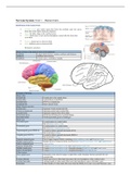
Cerebral cortex: grey matter region that forms the cerebrum outer rim; grows
faster than white matter during development
Gyrus: the folds; ridge on the cerebral cortex
Lobes: subdivision of each cerebral hemisphere, named after the bones that
cover them
Fissures: deepest groves between folds
Sulci: shallower grooves between folds
Division by structure
Major division of the brain (in terms of development)
Forebrain Largest brain division; contains cerebrum and thalamus
Midbrain Smallest brain division
Hindbrain Contains pons, cerebellum, medulla
Features of the brain
Major lobes
Frontal lobe Frontal pole to the central sulcus
Parietal lobe Central sulcus to the POCS
Occipital lobe POCS to occipital pole
Temporal lobe Below lateral fissure
Major fissures and sulci
Central sulci Separates frontal and parietal lobe
Parietal occipital sulci Separates parietal and occipital lobe
Lateral fissure Separates temporal lobes from others
Postcentral sulcus Divides postcentral gyrus to parietal lobule
Precentral sulcus Divides precentral gyrus to frontal gyri
Major gyri
Precentral gyrus Located anterior to central sulcus
Site of primary motor cortex
Postcentral gyrus Located posterior to central sulcus
Site of primary somatosensory cortex
Supramarginal gyrus (SMGLA) Located in inferior parietal lobe beside AGLA
Site involved in reading
Angular gyrus (AGLA) Located in inferior parietal lobe beside SMGLA
Site involved in writing
Superior temporal gyrus Part of upper temporal lobule containing primary auditory cortex and Wernicke’s area
Middle temporal gyrus Middle and lower temporal lobule, also known as temporal association cortex and supplementary
Inferior temporal gyrus visual cortices
Superior frontal gyrus Contains frontal association cortex and supplementary motor area
Middle frontal gyrus Contains frontal association cortex, alongside planning frontal eye field and Exner’s area
Inferior frontal gyrus Contains frontal association cortex, alongside planning Broca’s speech area
Other features
Preoccipital notch Indentation in the inferior margin occipital lobe; marks boundary between parietal lobe and
occipital lobe
Corpus callosum Thick band of nerve fibres that interconnect the two hemispheres of the cerebral cortex
Arcuate fasciculus Arching bundle of white matter fibre tracts that links Broca’s area to Wernicke’s area
Fasciculus Other bundles of white matter fibre tracts that link SMGLA to Exner’s area and Wernicke’s area
, Division by functional areas
Functions of the brain
Site Location Function
Primary visual cortex Posterior tip of median occipital lobe Receives visual information and is involved in visual
perception
Supplementary visual cortices Remaining region of occipital lobe Interpret visual information received
Primary auditory area Superior temporal gyrus Receives information for sound and is involved in auditory
perception
Wernicke’s speech area Superior temporal gyrus Interpret speech; also known as secondary language area
Supramarginal gyrus (SMGLA) Inferior parietal lobe beside AGLA Site involved in reading
Angular gyrus (AGLA) Inferior parietal lobe beside SMGLA Site involved in writing
Primary motor cortex Precentral gyrus Voluntary contractions of specific muscles on the opposite
side of the body; muscle represented unequally (e.g. more
cortical area devoted to fingers than to hip)
Primary somatosensory cortex Postcentral gyrus Receives nerve impulses for touch, pressure, vibration,
temperature etc.
Premotor cortex Anterior to primary motor cortex Planning or preparation for movement
Broca’s speech area Inferior frontal gyrus Produces speech; also known as primary language area
Frontal eye field Middle frontal gyrus (below Exner’s) Coordinate movement of visual perception (e.g. eye)
Exner’s area Middle frontal gyrus (above FEF) Coordinate movement of hands
Sensory homunculus: distorted map of the entire body in terms of
sensation where each point of the primary somatosensory cortex
receives impulses from a specific part of the body
Motor homunculus: distorted map of the entire body in terms of motor
where each point of the primary motor cortex receives is responsible of
movement of specific part of the body
Dominant hemisphere: the hemisphere where all the functional regions
related to speech and language are present (usually left)
Functions of the brain (association)
Region Location Involved in
Frontal association cortex Frontal lobe Intelligence
Personality
Behaviour
Mood
Cognitive function
Parietal association cortex Parietal lobe Spatial skills
3D recognition (shapes, faces, concepts, abstract perceptions)
Temporal association Temporal lobe Memory
cortex Mood
Aggression
Intelligence
Note that the specific details of the frontal association cortex are not clearly defined
Effects of lesions
Functional effects of lesions of the cerebral cortex
Type of aphasia Site of damage Consequences
Motor aphasia Broca’s area Person can interpret speech but cannot produce speech coherently
Connective aphasia Arcuate fasciculus Person can understand and interpret sound/speech and produce speech, but
has difficulty connecting the two
Sensory aphasia Wernicke’s area Person cannot interpret speech
,Non-dominant brain
Non-dominant hemisphere (right): all functional region related to speech and language present in only one hemisphere (left)
Functions of non-dominant hemisphere:
Non-verbal language (e.g. body language)
Emotional expression (e.g. tone of language)
Spatial skills (3D)
Conceptual understanding
Artistic and musical skills
Effects of injury in non-dominant hemisphere:
Loss of non-verbal language
Emotionless speech
Spatial disorientation
Inability to recognize familiar objects
Loss of musical appreciation
Damage to left brain that cause individual to lose ability of normal speech may still allow them to sing
, Nervous System: Unit 2 – Spinal cord
Central nervous system (CNS): part of the nervous system consisting of the brain and spinal cord
Spinal cord: thin tube of nervous tissue running through the vertebral column
Length: extends from the medulla oblongata of the hindbrain to the conus medullaris
Anatomy of the spinal cord
External anatomy of the spinal cord
Spinal nerves
Cervical nerves 8 pairs Innervates upper parts of the body (i.e.
head, shoulder, arms and fingers)
Thoracic nerves 12 pairs Innervates median-upper parts of the
body (i.e. chest)
Lumbar nerves 5 pairs Innervates anterior lower parts of the
body (i.e. anterior legs and feet)
Sacral nerves 5 pairs Innervates posterior lower parts of the
body (i.e. buttocks, posterior thigh,
lower leg and foot, reproductive organs,
foot)
Coccygeal nerves 1 pair Innervates around the tailbone
Other features
Cervical Enlargement of the grey matter that that contains the
enlargement neural machinery necessary to operate the upper
limbs (C4-1T)
Lumbar enlargement Enlargement of the grey matter that that contains the
neural machinery necessary to operate the lower
limbs (T9-T12)
Conus medullaris Cone shaped lower end of the spinal cord (L1-L2)
Cauda equina Bundle of spinal nerves and spinal nerve rootlets,
consisting of the L2 to the coccygeal nerve
Dermatome: an area of skin that is mainly supplied by a
single spinal nerve (homunculus representation of nerves)
Myotome: group of muscles that a single spinal nerve
innervates (homunculus representation of muscles)
Internal anatomy of the spinal cord and periphery (simple)
Grey matter Dark due to present of cell body
White matter Light due to absence of cell body
Dorsal Involved with sensory
Ventral Involved with motor
Dorsal root ganglion Cluster of neurons in a dorsal root of a
spinal nerve – contains sensory neurons
Note that unlike the brain where the grey matter is present at the outside, the grey matter is present inside of the spinal cord
Sensory (afferent) pathway
Sensory neurons are pseudounipolar: they contain an axon that has split into
two branches; one branch runs to the periphery and the other to the spinal cord
Information pathway:
Signal detected at periphery: touch/pressure signal is detected by the receptor
Dorsal root ganglia: signal arrives at the cell body at root ganglia
White column: the neuron involved is branched to the dorsal white matter (and to the brain)
Discriminative sensation information: ability to sense touch, and distinguish between two different senses of touch
Ability to discriminate: maximized in locations with most senses/receptors; more sensitive for fingers than foot
Specific to encapsulated receptors: such ability is specific to touch and pressure
2. Sensing pain and temperature:
Neurons involved: unmyelinated
Receptor type: not encapsulated (free nerve ending)
The benefits of buying summaries with Stuvia:
Guaranteed quality through customer reviews
Stuvia customers have reviewed more than 700,000 summaries. This how you know that you are buying the best documents.
Quick and easy check-out
You can quickly pay through credit card or Stuvia-credit for the summaries. There is no membership needed.
Focus on what matters
Your fellow students write the study notes themselves, which is why the documents are always reliable and up-to-date. This ensures you quickly get to the core!
Frequently asked questions
What do I get when I buy this document?
You get a PDF, available immediately after your purchase. The purchased document is accessible anytime, anywhere and indefinitely through your profile.
Satisfaction guarantee: how does it work?
Our satisfaction guarantee ensures that you always find a study document that suits you well. You fill out a form, and our customer service team takes care of the rest.
Who am I buying these notes from?
Stuvia is a marketplace, so you are not buying this document from us, but from seller ibdiplomamsg. Stuvia facilitates payment to the seller.
Will I be stuck with a subscription?
No, you only buy these notes for $24.89. You're not tied to anything after your purchase.