Proteins: Unit 2 – Buffers
pH: − log [ ]; describes the acidity/basicity of a solution
pKa: − log ; dissociation constant that describes the property of weak
acid side chain to lose a proton
Buffer: solution that resist pH changes; can maintain a nearly constant pH if
it is diluted, or if relatively small amounts of strong acids or bases are added
pH range in human blood: very tight range between 7.35 and 7.45
Blood in lung: tends towards a higher pH (less acidic)
Blood in tissues: tends towards a lower pH (more acidic;
promotes the release of oxygen at the tissue)
Systems that maintain human blood pH
System Timeframe Description
Respiratory Short term At higher pH, respiratory rate decreases,
system while at lower pH, respiratory rate increases
to remove CO2 from the system
Renal system Long term Regulate reabsorption of carbonic acid in the
tubule instead of secretion through urine
Chemical Immediate Small amount of acid/base does not
buffering systems dramatically change pH like water
Bicarbonate Dissolution of carbon dioxide in water
catalysed by carbonic anhydrase to carbonic
acid to dissociate into bicarbonate and H+
and vice versa
Proteins Contributes to buffering capacity via their
electrically-charged side chains or other
ionisable protein groups
Phosphates Dissolution of phosphates into its conjugate
base form and H+ and vice versa
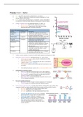
All these systems interplay to keep the blood pH tightly regulated (see diagram on right)
Bicarbonate system in the blood: equilibrium among CO2, H2CO3, HCO3-
Carbon dioxide transfer: carbon dioxide is transferred from body
tissue to blood, to RBC
Carbonic acid formation: carbon dioxide and water is converted to
carbonic acid by carbonic anhydrase
Buffer system: carbonic acid can dissociate into bicarbonate ion; act
as a buffer
Note that the bicarbonate system is the most dominant buffer system
pH regulation by respiratory system:
More CO2: more CO2 is produced due to active cell metabolism
Lower PH: more CO2 dissolved in blood lowers blood pH slightly
Brain signalling: receptors in the brain sense the drop in pH and
send nerve signals to increase breathing rates
Removable of CO2: increased breathing rate quickly removes more
CO2 to maintain homeostasis
Reason for multiple mechanisms:
Redundancy: act as a ‘backup’ to ensure maintenance of constant pH
Timing: systems act on different timing for different purposes
Forms of amino acids for proteins: each side chain will have a distinct pKa value, which, when
compared to the pH, can help to identify which form the amino acid is most likely found in
pH < pKa: equilibrium to the left; protonated (acidic) form
pH = pKa: both form exists in 50:50 mixture
pH > pKa: equilibrium to the right, deprotonated (basic) form
Aspartic and glutamic acid: low pKa; at standard
physiological pH, we expect these to be deprotonated
Lysine and arginine: high pKa; at standard
physiological pH, we expect these to be protonated
Histidine: this protein typically displays a wide pKa
range and may be protonated or deprotonated at
neutral pH depending on the environment the amino
acid is exposed to
, Bohr effect: affinity of oxygen to haemoglobin is inversely proportional to both
acidity and CO2 concentration
T state: deoxyhaemoglobin (lacks oxygen)
R state: oxyhaemoglobin (holds oxygen)
In lungs: pH = 7.4, and pH > pKa
Form: His146 most likely deprotonated
State: R state favoured (oxyhaemoglobin)
Oxygen: oxygen binding favoured; oxygen carried
In tissues: pH = 7.2 and pH < pKa
Form: His146 most likely protonated
State: T state favoured (deoxyhaemoglobin)
Oxygen: oxygen binding unfavoured; oxygen released
,Proteins: Unit 3 – Secondary structures
Secondary structure of a protein: describes the arrangement in space and the specific
hydrogen bonding of the peptide backbone
Characteristics: highly regular and repetitive
Types: α-helices, β-pleated sheets
Examples: wool and hair (keratin), silk (fibroin, spider silk)
Van der Waals interaction: weak interactions between all atoms that come close
enough to each other
Found in proteins: one of the types of interaction that hold a protein
together (tertiary); tightly packed for maximized contact
Strength: weaker than hydrogen bonds
Example: gecko can climb sheer surface through VdW interactions
Fibrous proteins
Fibrous protein: protein almost completely consisting of secondary structure
Repetitive: high regular; have repetitive structure and repetitive amino acid sequences
Strength: have mechanical and structural strength
Examples: wool and hair (keratin), silk (fibroin), collagen (skin)
Fibrous protein types
α-helix fibrous proteins β-sheet fibrous proteins
Key structure Helical protein (multiple S-S bonds) Sheet forming proteins
Trait Tough, insoluble with varying hardness and flexibility Soft, flexible
Example α-keratin of hair, feather, nail silk fibroin, spider silk
Β-sheet: a flexible sheet of proteins that can form barrels
Features: flexible, repetitive
Hydrogen bonding: every backbone N−H group donates a
hydrogen bond to the backbone C=O group of the amino acid of
a different polypeptide located opposite
Structure of silk:
Sheet structure: each polypeptide chain of repeating glycine and
alanine (or serine); all glycine is on one side, alanine on another
Hydrogen bonding: backbones of closely placed polypeptides
interact with each other such that hydrogen bonds are formed
Interdigitation: side chains fit in the gaps (e.g. alanine of one
polypeptide fits in the gap between two alanine of another)
Flexibility: the gap is not tight, allowing slight movement
Fibre direction: fibre direction is perpendicular to the polypeptide
α-helix: a right-hand spiral conformation (i.e. helix) of amino acids
Features: rigid, coiled and compact
Direction: right-handed
Amino acids per turn: 3.6 aa per turn
Hydrogen bonding: every backbone N−H group donates a
hydrogen bond to the backbone C=O group of the amino acid
located three or four residues earlier along the protein sequence
Disrupted by proline: proline’s side chain that interact with the
N (proline has no N-H) overall causes a ‘kink’ in the structure
Coiled-coil structure: structure in which multiple α-helices are coiled
together like the strands of a rope
Structure of hair:
Composition: repetitive sequence, ~14% cysteine
Helix structure: each polypeptide is a long α helix with a
globular head (repetitive)
Hydrophobic side: every 4th residue has a hydrophobic side
chain, such that when coiled, one side is hydrophobic
Coiled-coil structure: in hydrophilic environment, the helix
coils around each other (dimerization) such that the
hydrophobic part come together
Higher assembly: higher assembly to give wool fibres or hair
with filaments held together by H-bonds, ionic bonds,
disulphide bonds, and so forth
Fibre direction: fibre direction is alongside the polypeptide
Disulphide bond and curliness: more cysteine and more
disulphide bonds results in curlier hair
, Disulphide bonding in hair and perming:
Breaking of di-sulphide bond: di-sulphide
bonds are broken in hair
Curling: position of hair is changed (position
wanted to be fixed)
Di-sulphide bond reformation: neutralizer
treatment reforms the di-sulphide forms
Spidrion: main protein in a spider's dragline silk
Structure: mixture of beta-sheet nano-
crystalline regions and amorphous regions;
nanocrystals instead of whole beta sheets
makes structure stronger
Application: spider silk vests, clothing
Features: unique combination of tensile strength and extensibility
Strong: very strong; stronger than steel, similar to Kevlar
Stretchy: strength for functional purposes
Tough: high ability to absorbs energy and plastically deform without fracturing; than both steel or Kevlar
Peptide bonds: delocalized double bond that makes the peptide unit rigid and planar
The benefits of buying summaries with Stuvia:
Guaranteed quality through customer reviews
Stuvia customers have reviewed more than 700,000 summaries. This how you know that you are buying the best documents.
Quick and easy check-out
You can quickly pay through credit card or Stuvia-credit for the summaries. There is no membership needed.
Focus on what matters
Your fellow students write the study notes themselves, which is why the documents are always reliable and up-to-date. This ensures you quickly get to the core!
Frequently asked questions
What do I get when I buy this document?
You get a PDF, available immediately after your purchase. The purchased document is accessible anytime, anywhere and indefinitely through your profile.
Satisfaction guarantee: how does it work?
Our satisfaction guarantee ensures that you always find a study document that suits you well. You fill out a form, and our customer service team takes care of the rest.
Who am I buying these notes from?
Stuvia is a marketplace, so you are not buying this document from us, but from seller ibdiplomamsg. Stuvia facilitates payment to the seller.
Will I be stuck with a subscription?
No, you only buy these notes for $24.89. You're not tied to anything after your purchase.