On the diagnostics of lung tuberculosis most frequently are applied the following
X-ray methods:
1) x-ray;
2) radiography;
3) tomography;
4) fluorography.
Pulmonary tuberculosis is usually divided into primary and postprimary forms.
Forms of primary tuberculosis are
primary tubercular complex
bronchoadenitis.
Forms of post primary tuberculosis are
Disseminated lung tuberculosis
Focus lung tuberculosis
Infiltrative-pneumonic lung tuberculosis
Tuberculoma.
Cavernous lung tuberculosis
Fibrous cavernous lung tuberculosis.
Lung cirrhosis
Pleurisy.
PRIMARY TUBERCULAR COMPLEX
The classical primary complex consists of three basic components:
pulmonic, lymphadenitis and lymphangitis connecting them. However a phase of
infiltration passes before bipolarity becomes distinct on anterio-posterior
radiograph. An infiltration represents rather intensive opacity connected to a lung
root, sometimes it is deposited on the lung root. As a rule, infiltration is not
homogeneous. It’s borders are dim. The vessels and bronchi appear through
infiltration. The sizes of infiltrations are various and depend on a degree of lung’s
damage; they can be lobar, segmental and bronchopulmonary. The primary
complex is located in the top and middle lung segments more often. At dissolving
the sub-pleural localization of infiltration more distinctly is visible.
The primary complex has four stages of development:
I stage - pneumonic. On X-ray general view three components of a complex are
visible:
1) the focus in lung tissue by the size 2-4 cm. in diameter or more, of oval or
, 2
irregular form, various intensity (more often - average and even high), with an
indistinct, obscure contour; 2) the flow out to a root - lymphangitis, which is
defined as linear tension bars from focus to the root;
3) in a root - enlarged infiltrated lymphatic nodes. The root is represented to be
extended, its structure) is blurry, the intensity is increased. The contours outlining
lymphatic nodes, or are dim, or more precisely depict the increased nodes.
II stage - resorption. The size of the focus in lung tissue decreases, its intensity
raises, the contours become precise. The flow out to a root and infiltration of
lymphatic nodes decreases.
III stage - condensation. On a place of focus area remains with the size up to 1
cm, inside of it inclusions of calcinations appear as fine spots of sharp intensity.
Same spots of calcinations are noticeable and in lymphatic nodes of the lung root.
Thin tension bars are determined between the focus and the root.
IV stage - calcinations. The focus in lung tissue becomes even smaller, more
densely, of high intensity, with distinct contour, frequently rugged and rough.
Calcinations are intensified also in root lymphatic nodes. Calcinations in certain
cases are represented by solid, dense formations, in others - they have less
intensive shadows of inclusions, which testify about incomplete calcifications of
the focus and preservation of caseous regions in it.
At favorable course of primary tuberculosis complex with time calcification
increases up to ossification at the place of former caseosis located in peripheral
parts of lungs. This is Gohn's focus.
.
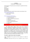
Radiographic picture of a primary tubercular complex (PC).
PC phase 1 - PC phase 2- PC phase 3 -
Normal infiltration resorption condensation
Outcome of
PC phase 4 - Gohn's
PC -
calcination focus
restoration
The benefits of buying summaries with Stuvia:
Guaranteed quality through customer reviews
Stuvia customers have reviewed more than 700,000 summaries. This how you know that you are buying the best documents.
Quick and easy check-out
You can quickly pay through credit card or Stuvia-credit for the summaries. There is no membership needed.
Focus on what matters
Your fellow students write the study notes themselves, which is why the documents are always reliable and up-to-date. This ensures you quickly get to the core!
Frequently asked questions
What do I get when I buy this document?
You get a PDF, available immediately after your purchase. The purchased document is accessible anytime, anywhere and indefinitely through your profile.
Satisfaction guarantee: how does it work?
Our satisfaction guarantee ensures that you always find a study document that suits you well. You fill out a form, and our customer service team takes care of the rest.
Who am I buying these notes from?
Stuvia is a marketplace, so you are not buying this document from us, but from seller serjmjl. Stuvia facilitates payment to the seller.
Will I be stuck with a subscription?
No, you only buy these notes for $7.49. You're not tied to anything after your purchase.