Surgery EOR Study Guide
GI/Nutritional (50%)
1) Abdominal Pain
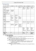
RUQ Epigastric LUQ Periumbilical RLQ Suprapubic LLQ
Colonic Colitis, Early Appendicitis, Appendicitis, Colitis,
Diverticulitis appendicitis colitis, colitis, diverticulitis,
diverticulitis, diverticulitis, IBD, IBS
IBD, IBS IBD, IBS
Biliary Cholecystitis, Cholecystitis,
cholelithiasis, cholelithiasis,
cholangitis cholangitis
Hepatic Abscess,
hepatitis, mass
Pulm PNA, embolus
Cardiac MI, Angina, MI,
pericarditis pericarditis
Vascular Aortic Aortic Aortic
dissection, dissection, dissection,
mesenteric mesenteric mesenteric
ischemia ischemia ischemia
Pancreat Mass, Mass,
ic pancreatitis pancreatitis
Renal Nephrolithiasis, Nephrolithias Nephrolithiasi Nephrolithiasi Nephrolithias
pyelonephritis is, s, s, is,
pyelonephriti pyelonephriti pyelnoephriti pyelonephriti
s s s, cystitis s
Gastric Esophagitis, Esophagitis, Esophagitis,
gastritis, PUD gastritis, gastritis,
PUD PUD, small
bowel mass,
obstruction
GYN Ectopic, Ectopic, Ectopic,
fibroids, fibroids, fibroids,
ovarian ovarian ovarian
mass, mass, mass,
torsion, PID, torsion, PID, torsion, PID,
endometriosi endometriosi endometriosi
s s s
Primary US CT CT w/ US CT w/ oral
test of contrast and IV
choice contrast
2) Acute/Chronic Cholecystitis
a. Acute Cholecystitis
i. Inflammation and infection of the gallbladder d/t obstruction of the cystic duct by
gallstones
ii. E. coli MC, Klebsiella & other gram (-) enteric organisms
iii. S/S: continuous RUQ or epigastric pain, may be precipitated by fatty foods or large
meals, may be assoc. with nausea, guarding & anorexia
iv. PE: fever (often low grade), enlarged, palpable GB
1. (+) Murphy’s sign – RUQ pain or inspiratory arrest w/ palpation of the GB
2. (+) Boas sign – referred pain to the right shoulder or subscapular area
(phrenic N. irritation)
v. Diagnosis: US = initial test of choice
1. Shows thickened or distended GB, pericholecystic fluid, sonographic Murphy’s
sign
, 2. CT – alternative to US and can detect complications
3. Labs – increased WBC (leukocytosis w/ left shift), increased bilirubin, increased
ALP, increased LFTs
4. HIDA scan = most accurate test – cholecystitis is present if no visualization of
GB
vi. Management:
1. NPO, IV fluids, antibiotics (s/a Ceftriaxone + Metronidazole) followed by
cholecystectomy (usually w/in 72h)
a. Laparoscopy preferred whenever possible
2. Cholecystectostomy (percutaneous drainage of GB) if pt is nonoperative
b. Acute Acalculous Cholecystitis
i. Acute necroinflammatory disease of the GB not due to gallstones
ii. Accounts for 10% of acute cholecystitis
iii. Pathophys: Gallbladder stasis & ischemia � local inflammatory reaction in the GB wall
� concentration of bile salts, GB distention, secondary infection, perforation or
necrosis of GB tissue
iv. RF: current hospitalization, critically ill pts
v. S/S: fever, leukocytosis, jaundice, sepsis, vague abdominal discomfort
vi. Diagnosis: based on clinical sx in the setting of supportive imaging & exclusion of
alternative dx
1. US = initial test of choice – distended GB w/ thickened walls and
pericholecystic fluid WITHOUT calcifications
2. CT abdomen w/contrast if diagnosis remains uncertain after US
3. HIDA scan performed if dx remains uncertain after CT scan
vii. Management: supportive – IV fluids, bowel rest, pain control, correction of
electrolytes, broad-spectrum ABX
c. Chronic Cholecystitis
i. Fibrosis & thickening of the GB d/t chronic inflammatory cell infiltration of the GB
evident on histopathology
ii. The presence of chronic cholecystitis does not correlate with symptoms
iii. Almost always assoc. with gallstones
3) Acute/Chronic Pancreatitis
a. Acute Pancreatitis
i. Acinar cell injury � intracellular activation of pancreatic enzymes � autodigestion of
pancreas
ii. Etiologies:
1. Gallstones (40%) & ETOH abuse (35%) = 2 MC causes
2. Meds: Thiazides, Protease inhibitors, Estrogens, Didanosine, Exenatide,
Valproic acid
3. Others: iatrogenic (ERCP), malignancy, scorpion sting, idiopathic, trauma, CF,
hypertriglyceridemia, hypercalcemia, infection, abdominal trauma/mumps in
children
iii. S/S:
1. Epigastric pain – constant, boring, often radiates to back or other quadrants,
exacerbated if supine or eating & relieved w/ leaning forward, sitting or fetal
position
2. N/V & fever common, shock in severe case
3. Necrotizing or Hemorrhagic: (+) Cullen’s sign (periumbilical ecchymosis) and
(+) Grey Turner sign (flank ecchymosis)
iv. PE: epigastric tenderness, tachycardia, decreased BS may be seen secondary to
adynamic ileus, dehydration or shock if severe
v. Diagnostic Criteria: 2 of the following 3
1. Acute onset of persistent, severe epigastric pain often radiating to the back
2. Elevation in serum lipase or amylase 3+x UNL
3. Characteristic findings of acute pancreatitis on imaging (CT, MRI, US)
a. No imaging required if pt meets the first 2 criteria
vi. Diagnostic Labs:
, 1. Increased amylase & lipase – best initial tests
a. Lipase more specific than amylase; levels don’t equal severity (not
specific)
b. ALT 3-fold increase is highly suggestive of gallstone pancreatitis
c. Hypocalcemia – necrotic fat binds to Ca++, lowering serum levels
(saponification)
d. Leukocytosis, elevated glucose, bilirubin & triglycerides
vii. Diagnostic Imaging:
1. Abdominal CT = imaging test of choice – also recommended in pts who fail to
improve or worsen after 48h to assess extent of necrosis
a. MRI is an alternative
2. Transabdominal US – recommended to assess for gallstones & bile duct
dilation
3. Abdominal XR – “sentinel loop” (localized ileus of a segment of small bowel in
the LUQ), Colon cutoff sign (abrupt collapse of the colon near the pancreas)
a. Pancreatic calcification is suggestive of chronic pancreatitis
4. MRCP useful to detect stones, stricture or tumor
5. Chest XR – left sided, exudative pleural effusion in moderate to severe cases
viii. Management:
1. 90% recover w/o complications in 3-7 days & require supportive measures
only – “rest the pancreas”
2. Supportive – NPO, high-volume IV fluid resuscitation, Analgesia
a. LR preferred – assoc. with decreased systemic inflammatory response
compared to NS
b. Analgesia – s/a Meperidine (Demerol)
3. Antibiotics – not routinely used; broad-spectrum abx (s/a Imipenem) used if
severe infected pancreatic necrosis seen (>30% necrosis on CT or MRI)
4. Ranson Criteria – used to determine prognosis (can also use APACHE score)
a. If score >/= 3, severe pancreatitis is likely. If < 3, severe pancreatitis is
unlikely.
b. Score 0-2 = 2% mortality, score 3-4 = 15% mortality, score 5-6 = 40%
mortality, score 7-8 = 100% mortality
Admission W/in 48h
Glucose >200 mg/dL Calcium <8.0 mg/dL
Age >55 years Hematocrit fall >10%
LDH >350 IU/L Oxygen PO2 <60 mmHg
AST >250 IU/dL BUN >5 mg/DL p IV fluids
WBC >16,000/mcL Base deficit >4 mEq/L
Sequestration of fluid >6L
b. Chronic Pancreatitis
i. Progressive inflammatory changes to the pancreas that lead to loss of pancreatic
endocrine & exocrine function
ii. Etiologies: ETOH abuse (MC), idiopathic, hypocalcemia, hyperlipidemia, islet cell
tumors, familial, trauma, iatrogenic
1. Gallstones not as significant as in acute
iii. S/S: triad of calcifications, steatorrhea & DM = hallmark but seen in only 1/3 of pts
1. Weight loss, epigastric and/or back pain may be atypical or completely absent
iv. Dx:
1. Amylase & lipase usually normal or mildly elevated
2. CT: calcification of the pancreas, often done in pts with acute pain to r/o other
causes of abdominal pain
3. Abdominal radiographs: calcified pancreas
, 4. Endoscopic US or MRCP
5. Pancreatic function testing: fecal elastase most sensitive & specific,
pancreatic stimulation w/ secretin & CCK usually not done
v. Management:
1. ETOH abstinence, pain control, low fat diet, vitamin supplementation
2. PO pancreatic enzyme replacement
3. Pancreatectomy only if retractable pain despite medical therapy
4) Anal Disease (Fissures, Abscess, Fistula)
a. Anal Fissure
i. Painful linear tear/crack in the distal anal canal
ii. Etiologies: low-fiber diets, passage of large hard stools, constipation or other anal
trauma
iii. S/S: severe painful rectal pain & bowel movements causing the pt to refrain from
defecating, bright red blood per rectum
iv. PE: longitudinal tear in the anoderm that usually extends no more proximally than the
dentate line
1. MC at the posterior midline (99% men, 90% women), skin tags seen in chronic
v. Management: Supportive = 1st line management – warm water Sitz baths, analgesics,
high fiber diet, ↑ water intake, stool softeners, laxatives & mineral oil
1. >80% resolve spontaneously
2. 2nd line tx: topical vasodilators – NTG (ADR HA & dizziness), Nifedipine
ointment
3. Botox injections to reduce spasm of internal sphincter (may be more effective
than topical dilators)
4. Surgery – s/a lateral internal sphincterectomy (reserved for refractory cases)
b. Anorectal Abscess & Fistula
i. Abscess: often results from bacterial infection of anal ducts or glands
1. Pathogens: Staph aureus (MC), E. coli, Bacteroides, Proteus, Streptococcus
2. Post. rectal wall = MC site
ii. Fistula = open tract b/w two epithelium lined-areas, seen esp. with deeper abscesses
iii. S/S:
1. Abscess: anorectal swelling, rectal pain that is worse w/
sitting/coughing/defecation, +/- fever; focal edema, induration & fluctuance on
exam.
a. Deeper abscesses may only be palpated on DRE or seen on imaging
studies
2. Fistula: may cause anal discharge & pain
iv. Management:
1. I&D = mainstay of tx
2. Followed by WASH – Warm-water cleansing, Analgesics, Sitz baths, High-fiber
diet
3. Abx not usually required in simple case
5) Anorexia
a. Screening for Eating Disorders: SCOFF questions
i. Do you make yourself Sick because you feel uncomfortably full?
ii. Do you worry you have lost Control over how much you eat?
iii. Have you recently lost more than One stone (14 lbs or 6.35 kg) in a 3-month period?
iv. Do you believe yourself to be Fat when others say you are too thin?
v. Would you say that Food dominates your life?
vi. “Yes” to 2+ questions = eating disorder
b. MC in females age 14-21 y/o (F:M 3:1, median onset = 18 y/o)
c. RF: female gender, child sexual abuse, OCD, childhood/parental obesity, substance abuse,
rigidi perfectionist, professions that emphasize thinness, athletes, homosexual men
d. Basic principle is that starvation induces protein & fat catabolism � loss of cellular volume
and atrophy in kidneys, brain, heart, liver, intestines & muscles
e. DSM-5 Criteria:


