The nervous system consists of neurons. There is an important difference between neurons and other parts of
the brain and that is if another part of the body breaks (bone/blood vessel), it can potentially be reattached,
whereas if an axon of the neuron is transect, the part that is disconnected from the cell body will always
degenerate and disappear. The axon is solely dependent on the cell body for proteins and energy. Whenever
the nervous system is damaged, even if those axons are reattached to the cell, they will degenerate and
disappear and they would have to regrow.
A nerve is a bundle of axons which consists of both afferent (sensory) and efferent (motor) axons. A nerve
consists of a bundle of (afferent and or efferent) axons surrounded by a connective tissue sheath, and is
located in the peripheral nervous system. Tracts and fascicles etc. consist of bundles of either afferent or
efferent axons, and are present in the central nervous system. A neuron usually consists of an (afferent)
dendritic tree, a cell body (soma), and an (efferent) axon that branches into axon terminals. The word
“neuron” however often refers to just the cell body. Do not confuse the words nerve, tract/fascicle, neuron
and axon!
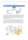
,Medical brain images are not mirror-images. Left and right in the images are co-oriented with the patient
standing in front of you. When it comes to how information in the brain passes to the body, this information
passes from the left to the right and vice versa. But depending on the type of sensory/motor information the
crossing happens at different stages.
Front and Back in the brain: we orient with respect to the long axis of the central nervous system, which
includes the spinal cord.
- Caudal = towards begin of the arrow.
- Rostral = towards point of the arrow.
- Ventral = towards the foreside of the arrow.
- Dorsal = towards the backside of the arrow.
Anatomical subdivision of the nervous system: The nervous system is subdivided anatomically into the central
nervous system and the peripheral nervous system. The central nervous system consists of the parts of the
nervous system encased by bones: the brain and spinal cord. Whatever is left outside belongs to the peripheral
nervous system, and consists of mainly just nerves (axons), either afferent or efferent nerves that can emerge
from different places. Cranial nerves considerably differ from each other, spinal nerves are all very similar.
Sometimes in the PNS neurons are found, and they are usually concentrated in groups which are called
ganglia.
Functional subdivision of the nervous system: This subdivision consists of the somatic and autonomic. The
somatic nervous system is what we have voluntary control over and is related to voluntary control
(somatomotor/somatosensory). The autonomic nervous system regulates our bodily functions without our
conscious awareness (viscerosensory: pain, pressure and visceromotor: sympathetic/parasympathetic).
Names derived from embryology
Sulcus: bordered on all sides by the neocortex
- Sulcus centralis cerebri (Fairly shallow; lined by neocortex only)
- Sulcus calcarinus (Deep; lined by neocortex only)
Fissure: bordered by other types of cortices
- Fissura longitudinalis cerebri (Very deep; lined by neocortex, allocortex and callosal body)
- Fissura lateralis cerebri (Deep and extensive; lined by neocortex and allocortex)
,NEUROCYTOLOGY: NEURONS AND OTHER CELLS
THE NEURON
The neuron is the most important cell in the nervous system:
Action potentials travel along axons and their presence is signalled to the next neuron at the synaps. At the
synapse a message is conveyed from one neuron to the next. Synapses impeatch on the dentrites where a
change of membrane potential will be created. At the axon hillock all the changes of the membrane potential
are integrated. If this sum reaches or surpases the threashold an action potential will emerge.
- All-or-none signal
- Unitary for each neuron (the same for each neuron, might be different between neurons)
- No attenuation
Saltatory conduction: an electrical impulse that skips from node to node down the full length of an axon,
speeding the arrival of the impulse at the nerve terminal in comparison with the slower continuous
progression of depolarization spreading down an unmyelinated axon.
, Synaptic transmission: Post synaptic potential differs from action potential as it is more variable, and it
diminishes the further away it gets.
Information along the neuron: it travels from the dendrites – cell body – axon – telodendrion – synaptic
terminals. From the dendrites to the cell body the information is carried by the post synaptic potential. At the
axon hillock the membrane potential is added and if it crosses the threshold an action potential is generated
which travels down from the axon to the synaptic terminals. At the synaptic terminals a neurotransmitter is
diffused into the next neuron to convey the information.
Interruption of the flow in information:
- Lesions at the afferent flow of information: sensory deficits, loss of sensory function
- Brain inside the cerebrum: complex changes of cognitive function
- Lesions in efferent flow: effector deficits, loss of somatomotor functions (paralysis, paresis), loss of
visceromotor functions (visceral dysfunctions).
The axons of afferent and efferent neurons are colocalized or located in close proximity and this is why lesions
usually may cause a mix of different sensorimotor symptoms. Thus, it is important to know how the afferent
and efferent systems merge together physically to be able to localize and understand a lesion.
The benefits of buying summaries with Stuvia:
Guaranteed quality through customer reviews
Stuvia customers have reviewed more than 700,000 summaries. This how you know that you are buying the best documents.
Quick and easy check-out
You can quickly pay through credit card or Stuvia-credit for the summaries. There is no membership needed.
Focus on what matters
Your fellow students write the study notes themselves, which is why the documents are always reliable and up-to-date. This ensures you quickly get to the core!
Frequently asked questions
What do I get when I buy this document?
You get a PDF, available immediately after your purchase. The purchased document is accessible anytime, anywhere and indefinitely through your profile.
Satisfaction guarantee: how does it work?
Our satisfaction guarantee ensures that you always find a study document that suits you well. You fill out a form, and our customer service team takes care of the rest.
Who am I buying these notes from?
Stuvia is a marketplace, so you are not buying this document from us, but from seller violinxris. Stuvia facilitates payment to the seller.
Will I be stuck with a subscription?
No, you only buy these notes for $7.47. You're not tied to anything after your purchase.